 Movie
Movie Controller

[English] 日本語
 Yorodumi
- PDB-6jnf: Cryo-EM structure of the translocator of the outer mitochondrial ... -
+ Open data

- Basic information
| Entry | Database: PDB / ID: 6jnf | ||||||||||||||||||
|---|---|---|---|---|---|---|---|---|---|---|---|---|---|---|---|---|---|---|---|
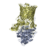
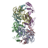
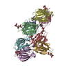
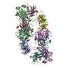
| Title | Cryo-EM structure of the translocator of the outer mitochondrial membrane | ||||||||||||||||||
 Components Components | (Mitochondrial import receptor subunit ...) x 5 | ||||||||||||||||||
 Keywords Keywords | TRANSLOCASE / alpha/beta translocator / membrane protein complex / Protein import / mitochondria | ||||||||||||||||||
| Function / homology |  Function and homology information Function and homology informationmitochondrial outer membrane translocase complex assembly / mitochondrial outer membrane translocase complex / protein insertion into mitochondrial outer membrane / protein transmembrane transport / : / porin activity / pore complex / protein import into mitochondrial matrix / transmembrane protein transporter activity / monoatomic ion transport ...mitochondrial outer membrane translocase complex assembly / mitochondrial outer membrane translocase complex / protein insertion into mitochondrial outer membrane / protein transmembrane transport / : / porin activity / pore complex / protein import into mitochondrial matrix / transmembrane protein transporter activity / monoatomic ion transport / intracellular protein transport / mitochondrial intermembrane space / mitochondrial outer membrane / mitochondrion / cytosol Similarity search - Function | ||||||||||||||||||
| Biological species |  | ||||||||||||||||||
| Method | ELECTRON MICROSCOPY / single particle reconstruction / cryo EM / Resolution: 3.81 Å | ||||||||||||||||||
 Authors Authors | Araiso, Y. / Tsutsumi, A. / Suzuki, J. / Yunoki, K. / Kawano, S. / Kikkawa, M. / Endo, T. | ||||||||||||||||||
| Funding support |  Japan, 5items Japan, 5items
| ||||||||||||||||||
 Citation Citation | Journal: Nature / Year: 2019 Title: Structure of the mitochondrial import gate reveals distinct preprotein paths. Authors: Yuhei Araiso / Akihisa Tsutsumi / Jian Qiu / Kenichiro Imai / Takuya Shiota / Jiyao Song / Caroline Lindau / Lena-Sophie Wenz / Haruka Sakaue / Kaori Yunoki / Shin Kawano / Junko Suzuki / ...Authors: Yuhei Araiso / Akihisa Tsutsumi / Jian Qiu / Kenichiro Imai / Takuya Shiota / Jiyao Song / Caroline Lindau / Lena-Sophie Wenz / Haruka Sakaue / Kaori Yunoki / Shin Kawano / Junko Suzuki / Marilena Wischnewski / Conny Schütze / Hirotaka Ariyama / Toshio Ando / Thomas Becker / Trevor Lithgow / Nils Wiedemann / Nikolaus Pfanner / Masahide Kikkawa / Toshiya Endo /     Abstract: The translocase of the outer mitochondrial membrane (TOM) is the main entry gate for proteins. Here we use cryo-electron microscopy to report the structure of the yeast TOM core complex at 3.8-Å ...The translocase of the outer mitochondrial membrane (TOM) is the main entry gate for proteins. Here we use cryo-electron microscopy to report the structure of the yeast TOM core complex at 3.8-Å resolution. The structure reveals the high-resolution architecture of the translocator consisting of two Tom40 β-barrel channels and α-helical transmembrane subunits, providing insight into critical features that are conserved in all eukaryotes. Each Tom40 β-barrel is surrounded by small TOM subunits, and tethered by two Tom22 subunits and one phospholipid. The N-terminal extension of Tom40 forms a helix inside the channel; mutational analysis reveals its dual role in early and late steps in the biogenesis of intermembrane-space proteins in cooperation with Tom5. Each Tom40 channel possesses two precursor exit sites. Tom22, Tom40 and Tom7 guide presequence-containing preproteins to the exit in the middle of the dimer, whereas Tom5 and the Tom40 N extension guide preproteins lacking a presequence to the exit at the periphery of the dimer. | ||||||||||||||||||
| History |
|
- Structure visualization
| Movie |
Movie viewer |
|---|---|
| Structure viewer | Molecule: MolmilJmol/JSmol |
- Downloads & links
-Download
| PDBx/mmCIF format | 6jnf.cif.gz | 185.1 KB | Display | PDBx/mmCIF format |
|---|---|---|---|---|
| PDB format | pdb6jnf.ent.gz | 138.1 KB | Display | PDB format |
| PDBx/mmJSON format | 6jnf.json.gz | Tree view | PDBx/mmJSON format | |
| Others |  Other downloads Other downloads |
-Validation report
| Arichive directory | https://data.pdbj.org/pub/pdb/validation_reports/jn/6jnfftp://data.pdbj.org/pub/pdb/validation_reports/jn/6jnf | HTTPS FTP |
|---|
-Related structure data
| Related structure data |  9851MC M: map data used to model this data C: citing same article ( |
|---|---|
| Similar structure data | |
| EM raw data | EMPIAR-10332 (Title: Cryo-EM structure of the translocator of the outer mitochondrial membrane Data size: 1.9 TB Data #1: Unaligned multi-frame micrographs of yeast TOM complex [micrographs - multiframe]) |


-Links
 PDBj
PDBj- Assembly
| Deposited unit | 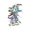
|
|---|---|
| 1 |
|
-Components
-Mitochondrial import receptor subunit ... , 5 types, 10 molecules AFBGCHDIEJ
| #1: Protein | Mass: 42071.141 Da / Num. of mol.: 2 Source method: isolated from a genetically manipulated source Source: (gene. exp.) #2: Protein | Mass: 6876.955 Da / Num. of mol.: 2 Source method: isolated from a genetically manipulated source Source: (gene. exp.) #3: Protein | Mass: 18481.139 Da / Num. of mol.: 2 Source method: isolated from a genetically manipulated source Source: (gene. exp.) #4: Protein/peptide | Mass: 5993.924 Da / Num. of mol.: 2 Source method: isolated from a genetically manipulated source Source: (gene. exp.) #5: Protein | Mass: 6410.460 Da / Num. of mol.: 2 Source method: isolated from a genetically manipulated source Source: (gene. exp.) |
|---|
-Non-polymers , 1 types, 1 molecules 
| #6: Chemical | ChemComp-46E / ( Mass: 635.853 Da / Num. of mol.: 1 / Source method: obtained synthetically / Formula: C33H66NO8P / Feature type: SUBJECT OF INVESTIGATION Mass: 635.853 Da / Num. of mol.: 1 / Source method: obtained synthetically / Formula: C33H66NO8P / Feature type: SUBJECT OF INVESTIGATION |
|---|
-Experimental details
-Experiment
| Experiment | Method: ELECTRON MICROSCOPY |
|---|---|
| EM experiment | Aggregation state: PARTICLE / 3D reconstruction method: single particle reconstruction |
- Sample preparation
| Component | Name: TOM complex / Type: COMPLEX / Entity ID: #1-#5 / Source: RECOMBINANT |
|---|---|
| Source (natural) | Organism: |
| Source (recombinant) | Organism: |
| Buffer solution | pH: 7.4 |
| Specimen | Conc.: 5.4 mg/ml / Embedding applied: NO / Shadowing applied: NO / Staining applied: NO / Vitrification applied: YES |
| Specimen support | Grid material: COPPER/RHODIUM / Grid mesh size: 300 divisions/in. / Grid type: Quantifoil, UltrAuFoil, R1.2/1.3 |
| Vitrification | Instrument: FEI VITROBOT MARK IV / Cryogen name: ETHANE / Humidity: 100 % / Chamber temperature: 279 K |
- Electron microscopy imaging
| Experimental equipment |  Model: Titan Krios / Image courtesy: FEI Company |
|---|---|
| Microscopy | Model: FEI TITAN KRIOS |
| Electron gun | Electron source:  FIELD EMISSION GUN / Accelerating voltage: 300 kV / Illumination mode: FLOOD BEAM FIELD EMISSION GUN / Accelerating voltage: 300 kV / Illumination mode: FLOOD BEAM |
| Electron lens | Mode: BRIGHT FIELD / Cs: 2.7 mm / C2 aperture diameter: 50 µm / Alignment procedure: COMA FREE |
| Specimen holder | Cryogen: NITROGEN / Specimen holder model: FEI TITAN KRIOS AUTOGRID HOLDER |
| Image recording | Electron dose: 50 e/Å2 / Film or detector model: FEI FALCON III (4k x 4k) |
- Processing
| Software |
| ||||||||||||||||||||||||||||||||
|---|---|---|---|---|---|---|---|---|---|---|---|---|---|---|---|---|---|---|---|---|---|---|---|---|---|---|---|---|---|---|---|---|---|
| EM software |
| ||||||||||||||||||||||||||||||||
| CTF correction | Type: PHASE FLIPPING AND AMPLITUDE CORRECTION | ||||||||||||||||||||||||||||||||
| Symmetry | Point symmetry: C2 (2 fold cyclic) | ||||||||||||||||||||||||||||||||
| 3D reconstruction | Resolution: 3.81 Å / Resolution method: FSC 0.143 CUT-OFF / Num. of particles: 124653 / Symmetry type: POINT | ||||||||||||||||||||||||||||||||
| Refinement | Stereochemistry target values: CDL v1.2 | ||||||||||||||||||||||||||||||||
| Refine LS restraints |
|
