[English] 日本語
 Yorodumi
- PDB-6jk7: Crystal structure of SpaE basal pilin from Lactobacillus rhamnosu... -
+ Open data
- Basic information
| Entry | Database: PDB / ID: 6jk7 | ||||||
|---|---|---|---|---|---|---|---|
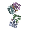
| Title | Crystal structure of SpaE basal pilin from Lactobacillus rhamnosus GG - Trigonal form | ||||||
 Components Components | Pilus assembly protein | ||||||
 Keywords Keywords | CELL ADHESION / basal pilins / SpaFED pilus / isopeptide bonds / pilus anchoring / surface proteins / probiotic / sortase | ||||||
| Function / homology |  Function and homology information Function and homology informationGram-positive pilin subunit D1, N-terminal / Gram-positive pilin subunit D1, N-terminal domain / Prealbumin-like fold domain / Prealbumin-like fold domain / Immunoglobulin-like fold / Immunoglobulins / Immunoglobulin-like / Sandwich / Mainly Beta Similarity search - Domain/homology | ||||||
| Biological species |  Lactobacillus rhamnosus (bacteria) Lactobacillus rhamnosus (bacteria) | ||||||
| Method |  X-RAY DIFFRACTION / X-RAY DIFFRACTION /  SYNCHROTRON / SYNCHROTRON /  MOLECULAR REPLACEMENT / Resolution: 3.204 Å MOLECULAR REPLACEMENT / Resolution: 3.204 Å | ||||||
 Authors Authors | Megta, A.K. / Mishra, A.K. / Palva, A. / von Ossowski, I. / Krishnan, V. | ||||||
| Funding support |  India, 1items India, 1items
| ||||||
 Citation Citation |  Journal: J.Struct.Biol. / Year: 2019 Journal: J.Struct.Biol. / Year: 2019Title: Crystal structure of basal pilin SpaE reveals the molecular basis of its incorporation in the lactobacillar SpaFED pilus. Authors: Megta, A.K. / Mishra, A.K. / Palva, A. / von Ossowski, I. / Krishnan, V. | ||||||
| History |
|
- Structure visualization
| Structure viewer | Molecule:  Molmil Molmil Jmol/JSmol Jmol/JSmol |
|---|
- Downloads & links
- Download
| PDBx/mmCIF format |  6jk7.cif.gz 6jk7.cif.gz | 137.3 KB | Display |  PDBx/mmCIF format PDBx/mmCIF format |
|---|---|---|---|---|
| PDB format |  pdb6jk7.ent.gz pdb6jk7.ent.gz | 105.4 KB | Display |  PDB format PDB format |
| PDBx/mmJSON format |  6jk7.json.gz 6jk7.json.gz | Tree view |  PDBx/mmJSON format PDBx/mmJSON format | |
| Others |  Other downloads Other downloads |
-Validation report
| Arichive directory |  https://data.pdbj.org/pub/pdb/validation_reports/jk/6jk7 https://data.pdbj.org/pub/pdb/validation_reports/jk/6jk7 ftp://data.pdbj.org/pub/pdb/validation_reports/jk/6jk7 ftp://data.pdbj.org/pub/pdb/validation_reports/jk/6jk7 | HTTPS FTP |
|---|
-Related structure data
| Related structure data |  6jbvSC 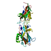 6jchC C: citing same article ( S: Starting model for refinement |
|---|---|
| Similar structure data |
- Links
Links
- Assembly
| Deposited unit | 
| ||||||||
|---|---|---|---|---|---|---|---|---|---|
| 1 |
| ||||||||
| Unit cell |
|
- Components
| #1: Protein | Mass: 44833.898 Da / Num. of mol.: 1 Source method: isolated from a genetically manipulated source Source: (gene. exp.)  Lactobacillus rhamnosus (strain ATCC 53103 / GG) (bacteria) Lactobacillus rhamnosus (strain ATCC 53103 / GG) (bacteria)Strain: ATCC 53103 / GG / Gene: DU507_12320 / Plasmid: pET28b(+) / Production host:  |
|---|---|
| Has protein modification | Y |
-Experimental details
-Experiment
| Experiment | Method:  X-RAY DIFFRACTION / Number of used crystals: 1 X-RAY DIFFRACTION / Number of used crystals: 1 |
|---|
- Sample preparation
| Crystal | Density Matthews: 3.22 Å3/Da / Density % sol: 61 % / Description: Three dimensional trigonal crystals |
|---|---|
| Crystal grow | Temperature: 295 K / Method: vapor diffusion, hanging drop / pH: 8.5 Details: 0.1 M bis-tris propane pH 8.5, 0.3 M sodium formate, 22% (w/v) PEG 3350 |
-Data collection
| Diffraction | Mean temperature: 100 K / Serial crystal experiment: N |
|---|---|
| Diffraction source | Source:  SYNCHROTRON / Site: SYNCHROTRON / Site:  ESRF ESRF  / Beamline: BM14 / Wavelength: 0.97887 Å / Beamline: BM14 / Wavelength: 0.97887 Å |
| Detector | Type: MARMOSAIC 225 mm CCD / Detector: CCD / Date: Sep 25, 2015 |
| Radiation | Protocol: SINGLE WAVELENGTH / Monochromatic (M) / Laue (L): M / Scattering type: x-ray |
| Radiation wavelength | Wavelength: 0.97887 Å / Relative weight: 1 |
| Reflection | Resolution: 3.204→65.023 Å / Num. obs: 9728 / % possible obs: 99.9 % / Redundancy: 5.6 % / CC1/2: 0.999 / Rmerge(I) obs: 0.064 / Rpim(I) all: 0.029 / Rrim(I) all: 0.071 / Net I/σ(I): 19.3 |
| Reflection shell | Resolution: 3.204→3.26 Å / Redundancy: 5.8 % / Rmerge(I) obs: 0.715 / Mean I/σ(I) obs: 2.5 / Num. unique obs: 495 / CC1/2: 0.76 / Rpim(I) all: 0.321 / Rrim(I) all: 0.785 / % possible all: 100 |
- Processing
| Software |
| |||||||||||||||||||||||||||||||||||||||||||||||||||||||||||||||||||||||||||
|---|---|---|---|---|---|---|---|---|---|---|---|---|---|---|---|---|---|---|---|---|---|---|---|---|---|---|---|---|---|---|---|---|---|---|---|---|---|---|---|---|---|---|---|---|---|---|---|---|---|---|---|---|---|---|---|---|---|---|---|---|---|---|---|---|---|---|---|---|---|---|---|---|---|---|---|---|
| Refinement | Method to determine structure:  MOLECULAR REPLACEMENT MOLECULAR REPLACEMENTStarting model: 6JBV Resolution: 3.204→52.495 Å / SU ML: 0.37 / Cross valid method: THROUGHOUT / σ(F): 1.35 / Phase error: 26.41
| |||||||||||||||||||||||||||||||||||||||||||||||||||||||||||||||||||||||||||
| Solvent computation | Shrinkage radii: 0.9 Å / VDW probe radii: 1.11 Å | |||||||||||||||||||||||||||||||||||||||||||||||||||||||||||||||||||||||||||
| Refinement step | Cycle: LAST / Resolution: 3.204→52.495 Å
| |||||||||||||||||||||||||||||||||||||||||||||||||||||||||||||||||||||||||||
| Refine LS restraints |
| |||||||||||||||||||||||||||||||||||||||||||||||||||||||||||||||||||||||||||
| LS refinement shell |
| |||||||||||||||||||||||||||||||||||||||||||||||||||||||||||||||||||||||||||
| Refinement TLS params. | Method: refined / Refine-ID: X-RAY DIFFRACTION
| |||||||||||||||||||||||||||||||||||||||||||||||||||||||||||||||||||||||||||
| Refinement TLS group |
|
 Movie
Movie Controller






 PDBj
PDBj