 Movie
Movie Controller

[English] 日本語
 Yorodumi
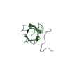
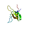
- PDB-6jje: Crystal structure of a two-quartet DNA mixed-parallel/antiparalle... -
+ Open data

- Basic information
| Entry | Database: PDB / ID: 6jje | ||||||||||||||||||||||||||||
|---|---|---|---|---|---|---|---|---|---|---|---|---|---|---|---|---|---|---|---|---|---|---|---|---|---|---|---|---|---|
| Title | Crystal structure of a two-quartet DNA mixed-parallel/antiparallel G-quadruplex (BrU) | ||||||||||||||||||||||||||||
 Components Components | DNA (5'-D(* Keywords KeywordsDNA / G-quadruplex / two-quartet / mixed-parallel/antiparallel | Function / homology | : / COBALT HEXAMMINE(III) / DNA / DNA (> 10) |  Function and homology information Function and homology informationBiological species |  Pseudorabies virus Ea Pseudorabies virus EaMethod |  X-RAY DIFFRACTION / SYNCHROTRON / MOLECULAR REPLACEMENT / Resolution: 1.378 Å X-RAY DIFFRACTION / SYNCHROTRON / MOLECULAR REPLACEMENT / Resolution: 1.378 Å  Authors AuthorsZhang, Y.S. / Wei, D.G. | Funding support | |  China, 1items China, 1items
 CitationJournal: To Be Published CitationJournal: To Be PublishedTitle: Crystal structure of a two-quartet DNA mixed-parallel/antiparallel G-quadruplex (BrU) Authors: Zhang, Y.S. / Parkinson, G.N. / Wagner, A. / Wei, D.G. History |
|
- Structure visualization
| Structure viewer | Molecule: MolmilJmol/JSmol |
|---|
- Downloads & links
-Download
| PDBx/mmCIF format | 6jje.cif.gz | 32.2 KB | Display | PDBx/mmCIF format |
|---|---|---|---|---|
| PDB format | pdb6jje.ent.gz | 20.9 KB | Display | PDB format |
| PDBx/mmJSON format | 6jje.json.gz | Tree view | PDBx/mmJSON format | |
| Others |  Other downloads Other downloads |
-Validation report
| Arichive directory | https://data.pdbj.org/pub/pdb/validation_reports/jj/6jjeftp://data.pdbj.org/pub/pdb/validation_reports/jj/6jje | HTTPS FTP |
|---|
-Related structure data
| Related structure data |  6jjfS S: Starting model for refinement |
|---|---|
| Similar structure data |


-Links
 PDBj
PDBj


- Assembly
| Deposited unit | 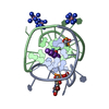
| ||||||||
|---|---|---|---|---|---|---|---|---|---|
| 1 |
| ||||||||
| Unit cell |
| ||||||||
| Components on special symmetry positions |
|
-Components
| #1: DNA chain | Mass: 4427.686 Da / Num. of mol.: 2 / Source method: obtained synthetically / Source: (synth.) Pseudorabies virus Ea#2: Chemical |   Mass: 39.098 Da / Num. of mol.: 3 / Source method: obtained synthetically / Formula: K / Feature type: SUBJECT OF INVESTIGATION Mass: 39.098 Da / Num. of mol.: 3 / Source method: obtained synthetically / Formula: K / Feature type: SUBJECT OF INVESTIGATION#3: Chemical |   Mass: 161.116 Da / Num. of mol.: 3 / Source method: obtained synthetically / Formula: CoH18N6 / Feature type: SUBJECT OF INVESTIGATION Mass: 161.116 Da / Num. of mol.: 3 / Source method: obtained synthetically / Formula: CoH18N6 / Feature type: SUBJECT OF INVESTIGATION#4: Chemical | ChemComp-NA / |   Mass: 22.990 Da / Num. of mol.: 1 / Source method: obtained synthetically / Formula: Na / Feature type: SUBJECT OF INVESTIGATION Mass: 22.990 Da / Num. of mol.: 1 / Source method: obtained synthetically / Formula: Na / Feature type: SUBJECT OF INVESTIGATION#5: Water | ChemComp-HOH / |  Mass: 18.015 Da / Num. of mol.: 95 / Source method: isolated from a natural source / Formula: H2O Mass: 18.015 Da / Num. of mol.: 95 / Source method: isolated from a natural source / Formula: H2O |
|---|
-Experimental details
-Experiment
| Experiment | Method: X-RAY DIFFRACTION / Number of used crystals: 1 |
|---|
- Sample preparation
| Crystal | Density Matthews: 2.13 Å3/Da / Density % sol: 42.27 % / Description: square |
|---|---|
| Crystal grow | Temperature: 283 K / Method: vapor diffusion, hanging drop Details: Potassium cacodylate,Sodium cacodylate,Potassium chloride,Sodium chloride,hexammine cobalt(III) chloride,MPD |
-Data collection
| Diffraction | Mean temperature: 100 K / Serial crystal experiment: N |
|---|---|
| Diffraction source | Source: SYNCHROTRON / Site: SSRF / Beamline: BL19U1 / Wavelength: 0.91944 Å |
| Detector | Type: DECTRIS PILATUS3 6M / Detector: PIXEL / Date: May 5, 2018 |
| Radiation | Protocol: SINGLE WAVELENGTH / Monochromatic (M) / Laue (L): M / Scattering type: x-ray |
| Radiation wavelength | Wavelength: 0.91944 Å / Relative weight: 1 |
| Reflection | Resolution: 1.378→50 Å / Num. obs: 15296 / % possible obs: 99.2 % / Redundancy: 6.5 % / Rmerge(I) obs: 0.063 / Net I/σ(I): 11.536 |
| Reflection shell | Resolution: 1.378→1.428 Å / Rmerge(I) obs: 0.289 / % possible all: 99.6 |
- Processing
| Software |
| ||||||||||||||||||||||||||||||||||||||||||
|---|---|---|---|---|---|---|---|---|---|---|---|---|---|---|---|---|---|---|---|---|---|---|---|---|---|---|---|---|---|---|---|---|---|---|---|---|---|---|---|---|---|---|---|
| Refinement | Method to determine structure: MOLECULAR REPLACEMENT Starting model: 6JJF Resolution: 1.378→31.49 Å / SU ML: 0.16 / Cross valid method: THROUGHOUT / σ(F): 1.42 / Phase error: 24.48
| ||||||||||||||||||||||||||||||||||||||||||
| Solvent computation | Shrinkage radii: 0.9 Å / VDW probe radii: 1.11 Å | ||||||||||||||||||||||||||||||||||||||||||
| Displacement parameters | Biso max: 84.41 Å2 / Biso mean: 19.3874 Å2 / Biso min: 6.24 Å2 | ||||||||||||||||||||||||||||||||||||||||||
| Refinement step | Cycle: final / Resolution: 1.378→31.49 Å
| ||||||||||||||||||||||||||||||||||||||||||
| LS refinement shell | Refine-ID: X-RAY DIFFRACTION / Rfactor Rfree error: 0 / Total num. of bins used: 5
|
