 Movie
Movie Controller

[English] 日本語
 Yorodumi
- PDB-6ji2: Crystal structure of archaeal ribosomal protein aP1, aPelota, and... -
+ Open data

- Basic information
| Entry | Database: PDB / ID: 6ji2 | ||||||
|---|---|---|---|---|---|---|---|
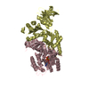
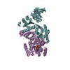
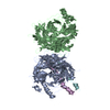
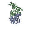
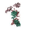
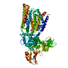
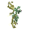
| Title | Crystal structure of archaeal ribosomal protein aP1, aPelota, and GTP-bound aEF1A complex | ||||||
 Components Components |
| ||||||
 Keywords Keywords | TRANSLATION / translation elongation ribosomal stalk | ||||||
| Function / homology |  Function and homology information Function and homology informationRNA surveillance / nuclear-transcribed mRNA catabolic process, no-go decay / nuclear-transcribed mRNA catabolic process, non-stop decay / ribosome disassembly / protein-synthesizing GTPase / nonfunctional rRNA decay / translational elongation / translation elongation factor activity / endonuclease activity / Hydrolases; Acting on ester bonds ...RNA surveillance / nuclear-transcribed mRNA catabolic process, no-go decay / nuclear-transcribed mRNA catabolic process, non-stop decay / ribosome disassembly / protein-synthesizing GTPase / nonfunctional rRNA decay / translational elongation / translation elongation factor activity / endonuclease activity / Hydrolases; Acting on ester bonds / structural constituent of ribosome / ribosome / ribonucleoprotein complex / hydrolase activity / GTPase activity / GTP binding / magnesium ion binding / metal ion binding / cytoplasm Similarity search - Function | ||||||
| Biological species |   Aeropyrum pernix K1 (archaea) Aeropyrum pernix K1 (archaea) | ||||||
| Method |  X-RAY DIFFRACTION / SYNCHROTRON / MOLECULAR REPLACEMENT / Resolution: 3 Å X-RAY DIFFRACTION / SYNCHROTRON / MOLECULAR REPLACEMENT / Resolution: 3 Å | ||||||
 Authors Authors | Maruyama, K. / Imai, H. / Kawamura, M. / Ishino, S. / Ishino, Y. / Ito, K. / Uchiumi, T. | ||||||
 Citation Citation | Journal: Sci Rep / Year: 2019 Title: Switch of the interactions between the ribosomal stalk and EF1A in the GTP- and GDP-bound conformations. Authors: Maruyama, K. / Imai, H. / Kawamura, M. / Ishino, S. / Ishino, Y. / Ito, K. / Uchiumi, T. | ||||||
| History |
|
- Structure visualization
| Structure viewer | Molecule: MolmilJmol/JSmol |
|---|
- Downloads & links
-Download
| PDBx/mmCIF format | 6ji2.cif.gz | 323.5 KB | Display | PDBx/mmCIF format |
|---|---|---|---|---|
| PDB format | pdb6ji2.ent.gz | 256.1 KB | Display | PDB format |
| PDBx/mmJSON format | 6ji2.json.gz | Tree view | PDBx/mmJSON format | |
| Others |  Other downloads Other downloads |
-Validation report
| Arichive directory | https://data.pdbj.org/pub/pdb/validation_reports/ji/6ji2ftp://data.pdbj.org/pub/pdb/validation_reports/ji/6ji2 | HTTPS FTP |
|---|
-Related structure data
| Related structure data |  3wxmS S: Starting model for refinement |
|---|---|
| Similar structure data |


-Links
 PDBj
PDBj


- Assembly
| Deposited unit | 
| ||||||||
|---|---|---|---|---|---|---|---|---|---|
| 1 | 
| ||||||||
| 2 | 
| ||||||||
| Unit cell |
|
-Components
-Protein , 2 types, 4 molecules AEBF
| #1: Protein | Mass: 49662.559 Da / Num. of mol.: 2 Source method: isolated from a genetically manipulated source Source: (gene. exp.) Aeropyrum pernix K1 (archaea) / Strain: K1 / Production host:  #2: Protein | Mass: 41470.602 Da / Num. of mol.: 2 Source method: isolated from a genetically manipulated source Source: (gene. exp.) Aeropyrum pernix K1 (archaea) / Strain: K1 / Production host: References: UniProt: Q9YAZ5, Hydrolases; Acting on ester bonds |
|---|
-Protein/peptide , 1 types, 1 molecules X
| #3: Protein/peptide | Mass: 1875.105 Da / Num. of mol.: 1 / Source method: obtained synthetically / Source: (synth.) Aeropyrum pernix K1 (archaea) / References: UniProt: Q9Y9W9*PLUS |
|---|
-Non-polymers , 4 types, 104 molecules 


| #4: Chemical |  Mass: 523.180 Da / Num. of mol.: 2 / Source method: obtained synthetically / Formula: C10H16N5O14P3 / Comment: GTP, energy-carrying molecule*YM Mass: 523.180 Da / Num. of mol.: 2 / Source method: obtained synthetically / Formula: C10H16N5O14P3 / Comment: GTP, energy-carrying molecule*YM#5: Chemical |  Mass: 24.305 Da / Num. of mol.: 2 / Source method: obtained synthetically / Formula: Mg Mass: 24.305 Da / Num. of mol.: 2 / Source method: obtained synthetically / Formula: Mg#6: Chemical |  Mass: 22.990 Da / Num. of mol.: 2 / Source method: obtained synthetically / Formula: Na Mass: 22.990 Da / Num. of mol.: 2 / Source method: obtained synthetically / Formula: Na#7: Water | ChemComp-HOH / | Mass: 18.015 Da / Num. of mol.: 98 / Source method: isolated from a natural source / Formula: H2O |
|---|
-Experimental details
-Experiment
| Experiment | Method: X-RAY DIFFRACTION / Number of used crystals: 1 |
|---|
- Sample preparation
| Crystal | Density Matthews: 2.98 Å3/Da / Density % sol: 58.74 % |
|---|---|
| Crystal grow | Temperature: 293 K / Method: vapor diffusion, sitting drop Details: 100 mM Tris-HCl, pH 8.0, 200 mM Li2SO4, 16% (w/v) PEG3350 |
-Data collection
| Diffraction | Mean temperature: 95 K / Serial crystal experiment: N |
|---|---|
| Diffraction source | Source: SYNCHROTRON / Site: Photon Factory  / Beamline: BL-5A / Wavelength: 1 Å / Beamline: BL-5A / Wavelength: 1 Å |
| Detector | Type: ADSC QUANTUM 315r / Detector: CCD / Date: May 12, 2017 |
| Radiation | Protocol: SINGLE WAVELENGTH / Monochromatic (M) / Laue (L): M / Scattering type: x-ray |
| Radiation wavelength | Wavelength: 1 Å / Relative weight: 1 |
| Reflection | Resolution: 3→106.42 Å / Num. obs: 41897 / % possible obs: 98.9 % / Redundancy: 3.9 % / Rmerge(I) obs: 0.07 / Net I/σ(I): 20.4 |
| Reflection shell | Resolution: 3→3.05 Å / Rmerge(I) obs: 0.564 |
- Processing
| Software |
| ||||||||||||||||||||||||||||||||||||||||||||||||||||||||||||||||||||||||||||||||||||||||||||||||||||||||||||||||||||||||||||||||||||||||||||||||||||||||||||||||||||||||||||||||||||||
|---|---|---|---|---|---|---|---|---|---|---|---|---|---|---|---|---|---|---|---|---|---|---|---|---|---|---|---|---|---|---|---|---|---|---|---|---|---|---|---|---|---|---|---|---|---|---|---|---|---|---|---|---|---|---|---|---|---|---|---|---|---|---|---|---|---|---|---|---|---|---|---|---|---|---|---|---|---|---|---|---|---|---|---|---|---|---|---|---|---|---|---|---|---|---|---|---|---|---|---|---|---|---|---|---|---|---|---|---|---|---|---|---|---|---|---|---|---|---|---|---|---|---|---|---|---|---|---|---|---|---|---|---|---|---|---|---|---|---|---|---|---|---|---|---|---|---|---|---|---|---|---|---|---|---|---|---|---|---|---|---|---|---|---|---|---|---|---|---|---|---|---|---|---|---|---|---|---|---|---|---|---|---|---|
| Refinement | Method to determine structure: MOLECULAR REPLACEMENT Starting model: 3WXM Resolution: 3→106.42 Å / Cor.coef. Fo:Fc: 0.94 / Cor.coef. Fo:Fc free: 0.873 / SU B: 21.804 / SU ML: 0.381 / Cross valid method: THROUGHOUT / ESU R Free: 0.482 / Details: HYDROGENS HAVE BEEN ADDED IN THE RIDING POSITIONS
| ||||||||||||||||||||||||||||||||||||||||||||||||||||||||||||||||||||||||||||||||||||||||||||||||||||||||||||||||||||||||||||||||||||||||||||||||||||||||||||||||||||||||||||||||||||||
| Solvent computation | Ion probe radii: 0.8 Å / Shrinkage radii: 0.8 Å / VDW probe radii: 1.2 Å | ||||||||||||||||||||||||||||||||||||||||||||||||||||||||||||||||||||||||||||||||||||||||||||||||||||||||||||||||||||||||||||||||||||||||||||||||||||||||||||||||||||||||||||||||||||||
| Displacement parameters | Biso mean: 82.131 Å2
| ||||||||||||||||||||||||||||||||||||||||||||||||||||||||||||||||||||||||||||||||||||||||||||||||||||||||||||||||||||||||||||||||||||||||||||||||||||||||||||||||||||||||||||||||||||||
| Refinement step | Cycle: 1 / Resolution: 3→106.42 Å
| ||||||||||||||||||||||||||||||||||||||||||||||||||||||||||||||||||||||||||||||||||||||||||||||||||||||||||||||||||||||||||||||||||||||||||||||||||||||||||||||||||||||||||||||||||||||
| Refine LS restraints |
|