 Movie
Movie Controller


+ Open data

- Basic information
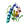
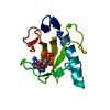
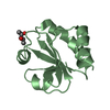
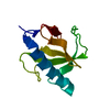
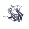
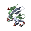
| Entry | Database: PDB / ID: 6jcc | ||||||
|---|---|---|---|---|---|---|---|
| Title | structure of a de novo protein D_1CY5_M1 | ||||||
 Components Components | Computational designed protein based on evolution | ||||||
 Keywords Keywords | DE NOVO PROTEIN / designed protein evolution crystal structure helix | ||||||
| Biological species | synthetic construct (others) | ||||||
| Method |  X-RAY DIFFRACTION / SYNCHROTRON / SAD / Resolution: 1.45 Å X-RAY DIFFRACTION / SYNCHROTRON / SAD / Resolution: 1.45 Å | ||||||
 Authors Authors | Meng, W. / Feng, T. | ||||||
| Funding support |  China, 1items China, 1items
| ||||||
 Citation Citation | Journal: To Be Published Title: structure of a computationally designed mutant protein D_1CY5_M1 Authors: Meng, W. / Haiyan, L. | ||||||
| History |
|
- Structure visualization
| Structure viewer | Molecule:  MolmilJmol/JSmol MolmilJmol/JSmol |
|---|
- Downloads & links
-Download
| PDBx/mmCIF format | 6jcc.cif.gz | 67.4 KB | Display | PDBx/mmCIF format |
|---|---|---|---|---|
| PDB format | pdb6jcc.ent.gz | 50.8 KB | Display | PDB format |
| PDBx/mmJSON format | 6jcc.json.gz | Tree view | PDBx/mmJSON format | |
| Others |  Other downloads Other downloads |
-Validation report
| Arichive directory | https://data.pdbj.org/pub/pdb/validation_reports/jc/6jccftp://data.pdbj.org/pub/pdb/validation_reports/jc/6jcc | HTTPS FTP |
|---|
-Related structure data
| Similar structure data |
|---|


-Links
 PDBj
PDBj
- Assembly
| Deposited unit | 
| ||||||||
|---|---|---|---|---|---|---|---|---|---|
| 1 |
| ||||||||
| Unit cell |
|
-Components
| #1: Protein | Mass: 11085.901 Da / Num. of mol.: 1 Source method: isolated from a genetically manipulated source Source: (gene. exp.) synthetic construct (others) Production host: |
|---|---|
| #2: Water | ChemComp-HOH /  Mass: 18.015 Da / Num. of mol.: 75 / Source method: isolated from a natural source / Formula: H2O Mass: 18.015 Da / Num. of mol.: 75 / Source method: isolated from a natural source / Formula: H2O |
-Experimental details
-Experiment
| Experiment | Method: X-RAY DIFFRACTION / Number of used crystals: 1 |
|---|
- Sample preparation
| Crystal | Density Matthews: 1.95 Å3/Da / Density % sol: 36.92 % |
|---|---|
| Crystal grow | Temperature: 285 K / Method: vapor diffusion, hanging drop Details: 0.1 M Sodium acetate trihydrate pH 4.6, 2.0 M Sodium formate |
-Data collection
| Diffraction | Mean temperature: 100 K / Serial crystal experiment: N |
|---|---|
| Diffraction source | Source: SYNCHROTRON / Site: SSRF / Beamline: BL17U / Wavelength: 0.979 Å |
| Detector | Type: ADSC QUANTUM 315r / Detector: CCD / Date: May 19, 2015 |
| Radiation | Protocol: SINGLE WAVELENGTH / Monochromatic (M) / Laue (L): M / Scattering type: x-ray |
| Radiation wavelength | Wavelength: 0.979 Å / Relative weight: 1 |
| Reflection | Resolution: 1.45→50 Å / Num. obs: 15784 / % possible obs: 98.8 % / Redundancy: 7.9 % / Biso Wilson estimate: 18.9 Å2 / Rmerge(I) obs: 0.061 / Rpim(I) all: 0.023 / Rrim(I) all: 0.065 / Net I/av σ(I): 4.588 / Net I/σ(I): 29.37 |
| Reflection shell | Resolution: 1.45→1.5 Å / Rmerge(I) obs: 0.422 / Num. unique obs: 1523 / Rpim(I) all: 0.159 |
- Processing
| Software |
| ||||||||||||||||||||
|---|---|---|---|---|---|---|---|---|---|---|---|---|---|---|---|---|---|---|---|---|---|
| Refinement | Method to determine structure: SAD / Resolution: 1.45→32.8 Å / Cross valid method: THROUGHOUT
| ||||||||||||||||||||
| Solvent computation | Bsol: 47.2 Å2 / ksol: 0.46 e/Å3 | ||||||||||||||||||||
| Displacement parameters | Biso mean: 16.63 Å2
| ||||||||||||||||||||
| Refinement step | Cycle: LAST / Resolution: 1.45→32.8 Å
| ||||||||||||||||||||
| LS refinement shell | Resolution: 1.451→1.489 Å
|
