 Movie
Movie Controller

[English] 日本語
 Yorodumi
- PDB-6jaz: Crystal structure of ABC transporter alpha-glycoside-binding muta... -
+ Open data

- Basic information
| Entry | Database: PDB / ID: 6jaz | |||||||||
|---|---|---|---|---|---|---|---|---|---|---|
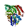
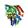
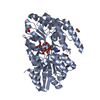
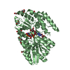
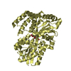
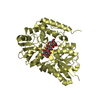
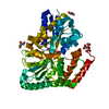
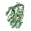
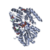
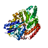
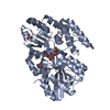
| Title | Crystal structure of ABC transporter alpha-glycoside-binding mutant protein W287F in complex with trehalose | |||||||||
 Components Components | ABC transporter, periplasmic substrate-binding protein | |||||||||
 Keywords Keywords | SUGAR BINDING PROTEIN / Carbohydrate-bindingsite / alpha-glycoside-binding protein / Ligand selection / Multi-substrate transporter / Sugar replacement / Venus Fly-trap mechanism | |||||||||
| Function / homology |  Function and homology information Function and homology information: / Bacterial extracellular solute-binding protein / Bacterial extracellular solute-binding protein / Bacterial extracellular solute-binding protein / Periplasmic binding protein-like II / D-Maltodextrin-Binding Protein; domain 2 / 3-Layer(aba) Sandwich / Alpha Beta Similarity search - Domain/homology | |||||||||
| Biological species |   Thermus thermophilus (bacteria) Thermus thermophilus (bacteria) | |||||||||
| Method |  X-RAY DIFFRACTION / MOLECULAR REPLACEMENT / molecular replacement / Resolution: 1.85 Å X-RAY DIFFRACTION / MOLECULAR REPLACEMENT / molecular replacement / Resolution: 1.85 Å | |||||||||
 Authors Authors | Kanaujia, S.P. / Chandravanshi, M. / Gogoi, P. | |||||||||
| Funding support |  India, 1items India, 1items
| |||||||||
 Citation Citation | Journal: Febs J. / Year: 2020 Title: Structural and thermodynamic correlation illuminates the selective transport mechanism of disaccharide alpha-glycosides through ABC transporter. Authors: Chandravanshi, M. / Gogoi, P. / Kanaujia, S.P. | |||||||||
| History |
|
- Structure visualization
| Structure viewer | Molecule: MolmilJmol/JSmol |
|---|
- Downloads & links
-Download
| PDBx/mmCIF format | 6jaz.cif.gz | 111.8 KB | Display | PDBx/mmCIF format |
|---|---|---|---|---|
| PDB format | pdb6jaz.ent.gz | 81.3 KB | Display | PDB format |
| PDBx/mmJSON format | 6jaz.json.gz | Tree view | PDBx/mmJSON format | |
| Others |  Other downloads Other downloads |
-Validation report
| Summary document | 6jaz_validation.pdf.gz | 885.5 KB | Display | wwPDB validaton report |
|---|---|---|---|---|
| Full document | 6jaz_full_validation.pdf.gz | 888.5 KB | Display | |
| Data in XML | 6jaz_validation.xml.gz | 22.7 KB | Display | |
| Data in CIF | 6jaz_validation.cif.gz | 35.3 KB | Display | |
| Arichive directory | https://data.pdbj.org/pub/pdb/validation_reports/ja/6jazftp://data.pdbj.org/pub/pdb/validation_reports/ja/6jaz | HTTPS FTP |
-Related structure data
| Related structure data |  6j9wSC  6j9yC 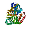 6jadC 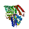 6jagC  6jahC 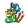 6jaiC 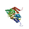 6jalC 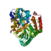 6jamC 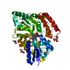 6janC  6jaoC 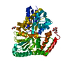 6japC  6jaqC 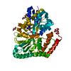 6jarC 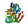 6jb0C  6jb4C  6jbaC 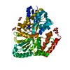 6jbbC 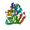 6jbeC S: Starting model for refinement C: citing same article ( |
|---|---|
| Similar structure data |


-Links
 PDBj
PDBj


- Assembly
| Deposited unit | 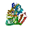
| ||||||||||||
|---|---|---|---|---|---|---|---|---|---|---|---|---|---|
| 1 |
| ||||||||||||
| Unit cell |
| ||||||||||||
| Components on special symmetry positions |
|
-Components
| #1: Protein | Mass: 46128.961 Da / Num. of mol.: 1 / Mutation: W287F Source method: isolated from a genetically manipulated source Source: (gene. exp.) Thermus thermophilus (strain HB8 / ATCC 27634 / DSM 579) (bacteria)Strain: HB8 / ATCC 27634 / DSM 579 / Gene: TTHA0356 / Plasmid: pET28a / Production host: |
|---|---|
| #2: Polysaccharide | alpha-D-glucopyranose-(1-1)-alpha-D-glucopyranose / trehalose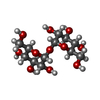  Source method: isolated from a genetically manipulated source Details: oligosaccharide with reducing-end-to-reducing-end glycosidic bond References: trehalose |
| #3: Chemical | ChemComp-CIT /   Mass: 192.124 Da / Num. of mol.: 1 / Source method: obtained synthetically / Formula: C6H8O7 Mass: 192.124 Da / Num. of mol.: 1 / Source method: obtained synthetically / Formula: C6H8O7 |
| #4: Chemical | ChemComp-EDO /   Mass: 62.068 Da / Num. of mol.: 1 / Source method: obtained synthetically / Formula: C2H6O2 Mass: 62.068 Da / Num. of mol.: 1 / Source method: obtained synthetically / Formula: C2H6O2 |
| #5: Water | ChemComp-HOH /  Mass: 18.015 Da / Num. of mol.: 539 / Source method: isolated from a natural source / Formula: H2O Mass: 18.015 Da / Num. of mol.: 539 / Source method: isolated from a natural source / Formula: H2O |
-Experimental details
-Experiment
| Experiment | Method: X-RAY DIFFRACTION / Number of used crystals: 1 |
|---|
- Sample preparation
| Crystal | Density Matthews: 2.92 Å3/Da / Density % sol: 57.85 % / Description: Tetragonal |
|---|---|
| Crystal grow | Temperature: 277 K / Method: microbatch / pH: 6.4 Details: 0.04 M Citric Acid, 0.06 M Bis-Tris Propne, 20% PEG 3350 |
-Data collection
| Diffraction | Mean temperature: 100 K / Serial crystal experiment: N |
|---|---|
| Diffraction source | Source: ROTATING ANODE / Type: RIGAKU MICROMAX-007 HF / Wavelength: 1.5418 Å |
| Detector | Type: RIGAKU RAXIS IV++ / Detector: IMAGE PLATE / Date: Feb 23, 2018 / Details: VariMax HF |
| Radiation | Protocol: SINGLE WAVELENGTH / Monochromatic (M) / Laue (L): M / Scattering type: x-ray |
| Radiation wavelength | Wavelength: 1.5418 Å / Relative weight: 1 |
| Reflection | Resolution: 1.85→73.59 Å / Num. obs: 46653 / % possible obs: 100 % / Redundancy: 14.8 % / CC1/2: 0.999 / Rmerge(I) obs: 0.092 / Rpim(I) all: 0.035 / Rrim(I) all: 0.098 / Net I/σ(I): 21.6 |
| Reflection shell | Resolution: 1.85→9.06 Å / Redundancy: 14.5 % / Rmerge(I) obs: 0.511 / Mean I/σ(I) obs: 5.8 / Num. unique obs: 2839 / CC1/2: 0.949 / Rpim(I) all: 0.196 / Rrim(I) all: 0.547 / % possible all: 99.9 |
-Phasing
| Phasing | Method: molecular replacement | |||||||||
|---|---|---|---|---|---|---|---|---|---|---|
| Phasing MR | Model details: Phaser MODE: MR_AUTO
|
- Processing
| Software |
| ||||||||||||||||||||||||||||||||||||||||||||||||||||||||||||
|---|---|---|---|---|---|---|---|---|---|---|---|---|---|---|---|---|---|---|---|---|---|---|---|---|---|---|---|---|---|---|---|---|---|---|---|---|---|---|---|---|---|---|---|---|---|---|---|---|---|---|---|---|---|---|---|---|---|---|---|---|---|
| Refinement | Method to determine structure: MOLECULAR REPLACEMENT Starting model: 6J9W Resolution: 1.85→73.59 Å / Cor.coef. Fo:Fc: 0.971 / Cor.coef. Fo:Fc free: 0.953 / SU B: 1.737 / SU ML: 0.053 / Cross valid method: THROUGHOUT / σ(F): 0 / ESU R: 0.091 / ESU R Free: 0.093 Details: HYDROGENS HAVE BEEN ADDED IN THE RIDING POSITIONS U VALUES : REFINED INDIVIDUALLY
| ||||||||||||||||||||||||||||||||||||||||||||||||||||||||||||
| Solvent computation | Ion probe radii: 0.8 Å / Shrinkage radii: 0.8 Å / VDW probe radii: 1.2 Å | ||||||||||||||||||||||||||||||||||||||||||||||||||||||||||||
| Displacement parameters | Biso max: 81.02 Å2 / Biso mean: 18.06 Å2 / Biso min: 8.07 Å2
| ||||||||||||||||||||||||||||||||||||||||||||||||||||||||||||
| Refinement step | Cycle: final / Resolution: 1.85→73.59 Å
| ||||||||||||||||||||||||||||||||||||||||||||||||||||||||||||
| Refine LS restraints |
| ||||||||||||||||||||||||||||||||||||||||||||||||||||||||||||
| LS refinement shell | Resolution: 1.85→1.898 Å / Rfactor Rfree error: 0 / Total num. of bins used: 20
|
