 Movie
Movie Controller

[English] 日本語
 Yorodumi
- PDB-6iz3: Structural basis for activity of TRIC counter-ion channels in cal... -
+ Open data

- Basic information
| Entry | Database: PDB / ID: 6iz3 | |||||||||
|---|---|---|---|---|---|---|---|---|---|---|
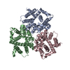
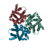
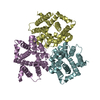
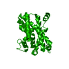
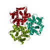
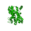
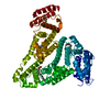
| Title | Structural basis for activity of TRIC counter-ion channels in calcium release | |||||||||
 Components Components | Trimeric intracellular cation channel type B-B | |||||||||
 Keywords Keywords | MEMBRANE PROTEIN | |||||||||
| Function / homology | TRIC channel / TRIC channel / regulation of release of sequestered calcium ion into cytosol / potassium channel activity / endoplasmic reticulum membrane / endoplasmic reticulum / identical protein binding / Trimeric intracellular cation channel type B-B Function and homology information Function and homology information | |||||||||
| Biological species | ||||||||||
| Method |  X-RAY DIFFRACTION / SYNCHROTRON / MOLECULAR REPLACEMENT / Resolution: 3.791 Å X-RAY DIFFRACTION / SYNCHROTRON / MOLECULAR REPLACEMENT / Resolution: 3.791 Å | |||||||||
 Authors Authors | Zeng, Y. / Wang, X.H. / Su, M. / Chen, Y.H. | |||||||||
| Funding support |  China, 2items China, 2items
| |||||||||
 Citation Citation | Journal: Proc.Natl.Acad.Sci.USA / Year: 2019 Title: Structural basis for activity of TRIC counter-ion channels in calcium release. Authors: Wang, X.H. / Su, M. / Gao, F. / Xie, W. / Zeng, Y. / Li, D.L. / Liu, X.L. / Zhao, H. / Qin, L. / Li, F. / Liu, Q. / Clarke, O.B. / Lam, S.M. / Shui, G.H. / Hendrickson, W.A. / Chen, Y.H. | |||||||||
| History |
|
- Structure visualization
| Structure viewer | Molecule: MolmilJmol/JSmol |
|---|
- Downloads & links
-Download
| PDBx/mmCIF format | 6iz3.cif.gz | 187.5 KB | Display | PDBx/mmCIF format |
|---|---|---|---|---|
| PDB format | pdb6iz3.ent.gz | 148.7 KB | Display | PDB format |
| PDBx/mmJSON format | 6iz3.json.gz | Tree view | PDBx/mmJSON format | |
| Others |  Other downloads Other downloads |
-Validation report
| Summary document | 6iz3_validation.pdf.gz | 478.4 KB | Display | wwPDB validaton report |
|---|---|---|---|---|
| Full document | 6iz3_full_validation.pdf.gz | 489.1 KB | Display | |
| Data in XML | 6iz3_validation.xml.gz | 30.8 KB | Display | |
| Data in CIF | 6iz3_validation.cif.gz | 40.9 KB | Display | |
| Arichive directory | https://data.pdbj.org/pub/pdb/validation_reports/iz/6iz3ftp://data.pdbj.org/pub/pdb/validation_reports/iz/6iz3 | HTTPS FTP |
-Related structure data
| Related structure data |  6iyuC  6iyxC  6iyzC  6iz0C 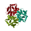 6iz1C  6iz4C  6iz5SC  6iz6C  6izfC S: Starting model for refinement C: citing same article ( |
|---|---|
| Similar structure data |


-Links
 PDBj
PDBj- Assembly
| Deposited unit | 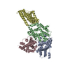
| ||||||||
|---|---|---|---|---|---|---|---|---|---|
| 1 | 
| ||||||||
| 2 | 
| ||||||||
| Unit cell |
|
-Components
| #1: Protein | Mass: 35463.840 Da / Num. of mol.: 4 Source method: isolated from a genetically manipulated source Source: (gene. exp.)  |
|---|
-Experimental details
-Experiment
| Experiment | Method: X-RAY DIFFRACTION / Number of used crystals: 1 |
|---|
- Sample preparation
| Crystal | Density Matthews: 4.32 Å3/Da / Density % sol: 71.56 % |
|---|---|
| Crystal grow | Temperature: 278 K / Method: vapor diffusion, sitting drop / Details: PEG 4000, KNO3 K-Citrate PH6.5 |
-Data collection
| Diffraction | Mean temperature: 100 K / Serial crystal experiment: N | ||||||||||||||||||||||||
|---|---|---|---|---|---|---|---|---|---|---|---|---|---|---|---|---|---|---|---|---|---|---|---|---|---|
| Diffraction source | Source: SYNCHROTRON / Site: APS  / Beamline: 24-ID-C / Wavelength: 1.7712 Å / Beamline: 24-ID-C / Wavelength: 1.7712 Å | ||||||||||||||||||||||||
| Detector | Type: DECTRIS PILATUS 300K / Detector: PIXEL / Date: Feb 21, 2017 | ||||||||||||||||||||||||
| Radiation | Protocol: SINGLE WAVELENGTH / Monochromatic (M) / Laue (L): M / Scattering type: x-ray | ||||||||||||||||||||||||
| Radiation wavelength | Wavelength: 1.7712 Å / Relative weight: 1 | ||||||||||||||||||||||||
| Reflection | Resolution: 3.78→47.85 Å / Num. obs: 24627 / % possible obs: 99.3 % / Redundancy: 37.5 % / CC1/2: 0.999 / Rmerge(I) obs: 0.237 / Rpim(I) all: 0.039 / Rrim(I) all: 0.241 / Net I/σ(I): 9.8 | ||||||||||||||||||||||||
| Reflection shell | Diffraction-ID: 1
|
- Processing
| Software |
| |||||||||||||||||||||||||||||||||||||||||||||||||||||||||||||||
|---|---|---|---|---|---|---|---|---|---|---|---|---|---|---|---|---|---|---|---|---|---|---|---|---|---|---|---|---|---|---|---|---|---|---|---|---|---|---|---|---|---|---|---|---|---|---|---|---|---|---|---|---|---|---|---|---|---|---|---|---|---|---|---|---|
| Refinement | Method to determine structure: MOLECULAR REPLACEMENT Starting model: 6IZ5 Resolution: 3.791→47.845 Å / SU ML: 0.77 / Cross valid method: THROUGHOUT / σ(F): 1.33 / Phase error: 42.43
| |||||||||||||||||||||||||||||||||||||||||||||||||||||||||||||||
| Solvent computation | Shrinkage radii: 0.9 Å / VDW probe radii: 1.11 Å | |||||||||||||||||||||||||||||||||||||||||||||||||||||||||||||||
| Displacement parameters | Biso max: 311.66 Å2 / Biso mean: 200.5016 Å2 / Biso min: 127.09 Å2 | |||||||||||||||||||||||||||||||||||||||||||||||||||||||||||||||
| Refinement step | Cycle: final / Resolution: 3.791→47.845 Å
| |||||||||||||||||||||||||||||||||||||||||||||||||||||||||||||||
| LS refinement shell | Refine-ID: X-RAY DIFFRACTION / Rfactor Rfree error: 0 / Total num. of bins used: 8
|
