 Movie
Movie Controller

[English] 日本語
 Yorodumi
- PDB-6iyw: Crystal sturucture of L,D-transpeptidase LdtMt2 from Mycobacteriu... -
+ Open data

- Basic information
| Entry | Database: PDB / ID: 6iyw | ||||||
|---|---|---|---|---|---|---|---|
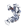
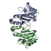
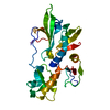
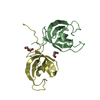
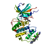
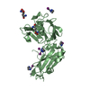
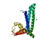
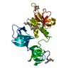
| Title | Crystal sturucture of L,D-transpeptidase LdtMt2 from Mycobacterium tuberculosis in complex with Imipenem adduct | ||||||
 Components Components | L,D-transpeptidase 2 | ||||||
 Keywords Keywords | TRANSFERASE / LD-TRANSPEPTIDASE / PEPTIDOGLYCAN SYNTHESIS ENZYME / beta-lactam binding | ||||||
| Function / homology |  Function and homology information Function and homology informationpeptidoglycan-protein cross-linking / peptidoglycan L,D-transpeptidase activity / Transferases; Acyltransferases; Aminoacyltransferases / acyltransferase activity / cell wall organization / regulation of cell shape / metal ion binding / plasma membrane Similarity search - Function | ||||||
| Biological species |   Mycobacterium tuberculosis (bacteria) Mycobacterium tuberculosis (bacteria) | ||||||
| Method |  X-RAY DIFFRACTION / SYNCHROTRON / MOLECULAR REPLACEMENT / Resolution: 1.6 Å X-RAY DIFFRACTION / SYNCHROTRON / MOLECULAR REPLACEMENT / Resolution: 1.6 Å | ||||||
 Authors Authors | Li, D.F. / Zhao, F. / Wang, D.C. | ||||||
| Funding support |  China, 1items China, 1items
| ||||||
 Citation Citation | Journal: Biochem. Biophys. Res. Commun. / Year: 2019 Title: The 1-beta-methyl group confers a lower affinity of l,d-transpeptidase LdtMt2 for ertapenem than for imipenem. Authors: Zhao, F. / Hou, Y.J. / Zhang, Y. / Wang, D.C. / Li, D.F. | ||||||
| History |
|
- Structure visualization
| Structure viewer | Molecule: MolmilJmol/JSmol |
|---|
- Downloads & links
-Download
| PDBx/mmCIF format | 6iyw.cif.gz | 357.6 KB | Display | PDBx/mmCIF format |
|---|---|---|---|---|
| PDB format | pdb6iyw.ent.gz | 289 KB | Display | PDB format |
| PDBx/mmJSON format | 6iyw.json.gz | Tree view | PDBx/mmJSON format | |
| Others |  Other downloads Other downloads |
-Validation report
| Arichive directory | https://data.pdbj.org/pub/pdb/validation_reports/iy/6iywftp://data.pdbj.org/pub/pdb/validation_reports/iy/6iyw | HTTPS FTP |
|---|
-Related structure data
| Related structure data |  6iyvC  3vypS S: Starting model for refinement C: citing same article ( |
|---|---|
| Similar structure data |


-Links
 PDBj
PDBj


- Assembly
| Deposited unit | 
| ||||||||
|---|---|---|---|---|---|---|---|---|---|
| 1 | 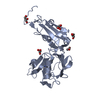
| ||||||||
| 2 | 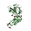
| ||||||||
| 3 | 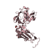
| ||||||||
| 4 | 
| ||||||||
| 5 | 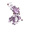
| ||||||||
| 6 | 
| ||||||||
| Unit cell |
|
-Components
| #1: Protein | Mass: 29552.693 Da / Num. of mol.: 6 Source method: isolated from a genetically manipulated source Source: (gene. exp.) Mycobacterium tuberculosis (strain ATCC 25618 / H37Rv) (bacteria)Strain: ATCC 25618 / H37Rv / Gene: ldtB, lppS, Rv2518c, RVBD_2518c, P425_02624 / Plasmid: pMCSG7 / Production host: References: UniProt: I6Y9J2, Transferases; Acyltransferases; Aminoacyltransferases #2: Chemical | ChemComp-IM2 / (   Mass: 301.362 Da / Num. of mol.: 6 / Source method: obtained synthetically / Formula: C12H19N3O4S / Comment: antibiotic*YM Mass: 301.362 Da / Num. of mol.: 6 / Source method: obtained synthetically / Formula: C12H19N3O4S / Comment: antibiotic*YM#3: Chemical | ChemComp-GOL /   Mass: 92.094 Da / Num. of mol.: 24 / Source method: obtained synthetically / Formula: C3H8O3 Mass: 92.094 Da / Num. of mol.: 24 / Source method: obtained synthetically / Formula: C3H8O3#4: Water | ChemComp-HOH / |  Mass: 18.015 Da / Num. of mol.: 1642 / Source method: isolated from a natural source / Formula: H2O Mass: 18.015 Da / Num. of mol.: 1642 / Source method: isolated from a natural source / Formula: H2OHas protein modification | Y | |
|---|
-Experimental details
-Experiment
| Experiment | Method: X-RAY DIFFRACTION / Number of used crystals: 1 |
|---|
- Sample preparation
| Crystal | Density Matthews: 2.19 Å3/Da / Density % sol: 43.84 % |
|---|---|
| Crystal grow | Temperature: 293 K / Method: vapor diffusion, sitting drop / pH: 6.5 / Details: NaCl, HEPES, pH 6.5, PEG 3350 |
-Data collection
| Diffraction | Mean temperature: 90 K |
|---|---|
| Diffraction source | Source: SYNCHROTRON / Site: SSRF / Beamline: BL19U1 / Wavelength: 0.9789 Å |
| Detector | Type: DECTRIS PILATUS3 6M / Detector: PIXEL / Date: Apr 28, 2018 |
| Radiation | Protocol: SINGLE WAVELENGTH / Monochromatic (M) / Laue (L): M / Scattering type: x-ray |
| Radiation wavelength | Wavelength: 0.9789 Å / Relative weight: 1 |
| Reflection | Resolution: 1.6→50 Å / Num. obs: 201419 / % possible obs: 98.8 % / Redundancy: 6.78 % / CC1/2: 0.998 / Rmerge(I) obs: 0.077 / Rrim(I) all: 0.084 / Net I/σ(I): 12.54 |
| Reflection shell | Resolution: 1.6→1.64 Å / Redundancy: 7.02 % / Rmerge(I) obs: 0.727 / Mean I/σ(I) obs: 2.79 / Num. unique obs: 14762 / CC1/2: 0.891 / % possible all: 98.1 |
- Processing
| Software |
| |||||||||||||||||||||||||||||||||||||||||||||||||||||||||||||||||||||||||||||||||||||||||||||||||||||||||
|---|---|---|---|---|---|---|---|---|---|---|---|---|---|---|---|---|---|---|---|---|---|---|---|---|---|---|---|---|---|---|---|---|---|---|---|---|---|---|---|---|---|---|---|---|---|---|---|---|---|---|---|---|---|---|---|---|---|---|---|---|---|---|---|---|---|---|---|---|---|---|---|---|---|---|---|---|---|---|---|---|---|---|---|---|---|---|---|---|---|---|---|---|---|---|---|---|---|---|---|---|---|---|---|---|---|---|
| Refinement | Method to determine structure: MOLECULAR REPLACEMENT Starting model: 3VYP Resolution: 1.6→46.414 Å / SU ML: 0.18 / Cross valid method: FREE R-VALUE / σ(F): 1.34 / Phase error: 24.46
| |||||||||||||||||||||||||||||||||||||||||||||||||||||||||||||||||||||||||||||||||||||||||||||||||||||||||
| Solvent computation | Shrinkage radii: 0.9 Å / VDW probe radii: 1.11 Å | |||||||||||||||||||||||||||||||||||||||||||||||||||||||||||||||||||||||||||||||||||||||||||||||||||||||||
| Refinement step | Cycle: LAST / Resolution: 1.6→46.414 Å
| |||||||||||||||||||||||||||||||||||||||||||||||||||||||||||||||||||||||||||||||||||||||||||||||||||||||||
| Refine LS restraints |
| |||||||||||||||||||||||||||||||||||||||||||||||||||||||||||||||||||||||||||||||||||||||||||||||||||||||||
| LS refinement shell |
|
