- PDB-3doa: The crystal structure of the fibrinogen binding protein from Stap... -
+
Open data
ID or keywords:
Loading...
-
Basic information
Entry
Database: PDB / ID: 3doa
Title
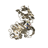
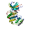
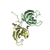
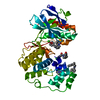
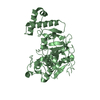
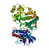
The crystal structure of the fibrinogen binding protein from Staphylococcus aureus
Components
Fibrinogen binding protein
Keywords
PROTEIN BINDING / The fibrinogen binding protein / structural genomics / MCSG. / PSI-2 / Protein Structure Initiative / Midwest Center for Structural Genomics
Function / homology
Function and homology information
RQC complex / ribosomal large subunit binding / rescue of stalled cytosolic ribosome / tRNA binding / rRNA binding Similarity search - Function
fibrinogen binding protein from staphylococcus aureus domain like / ibrinogen binding protein from staphylococcus aureus fold / ibrinogen binding protein from staphylococcus aureus domain / Ribonuclease HI; Chain A / Rqc2 homolog RqcH, bacterial / : / NFACT, RNA-binding domain / NFACT protein RNA binding domain / NFACT N-terminal and middle domains / Helicase, Ruva Protein; domain 3 - #50 ...fibrinogen binding protein from staphylococcus aureus domain like / ibrinogen binding protein from staphylococcus aureus fold / ibrinogen binding protein from staphylococcus aureus domain / Ribonuclease HI; Chain A / Rqc2 homolog RqcH, bacterial / : / NFACT, RNA-binding domain / NFACT protein RNA binding domain / NFACT N-terminal and middle domains / Helicase, Ruva Protein; domain 3 - #50 / Helicase, Ruva Protein; domain 3 / Roll / Orthogonal Bundle / 3-Layer(aba) Sandwich / Mainly Beta / Mainly Alpha / Alpha Beta Similarity search - Domain/homology
Resolution: 2.81→2.88 Å / Redundancy: 9.9 % / Mean I/σ(I) obs: 1.7 / Rsym value: 0.639 / % possible all: 88.09
-
Processing
Software
Name
Version
Classification
REFMAC
5.2.0019
refinement
SBC-Collect
datacollection
HKL-3000
datareduction
HKL-3000
datascaling
MLPHARE
phasing
Refinement
Method to determine structure: SAD / Resolution: 2.81→87.04 Å / Cor.coef. Fo:Fc: 0.953 / Cor.coef. Fo:Fc free: 0.891 / SU B: 29.329 / SU ML: 0.269 / TLS residual ADP flag: LIKELY RESIDUAL / Cross valid method: THROUGHOUT / ESU R: 0.633 / ESU R Free: 0.357 Stereochemistry target values: MAXIMUM LIKELIHOOD WITH PHASES Details: HYDROGENS HAVE BEEN ADDED IN THE RIDING POSITIONS
Rfactor
Num. reflection
% reflection
Selection details
Rfree
0.27048
530
4.8 %
RANDOM
Rwork
0.1872
-
-
-
obs
0.1908
10541
98.87 %
-
all
-
10661
-
-
Solvent computation
Ion probe radii: 0.8 Å / Shrinkage radii: 0.8 Å / VDW probe radii: 1.2 Å / Solvent model: MASK
Displacement parameters
Biso mean: 48.353 Å2
Baniso -1
Baniso -2
Baniso -3
1-
1.32 Å2
0 Å2
0 Å2
2-
-
1.32 Å2
0 Å2
3-
-
-
-2.64 Å2
Refinement step
Cycle: LAST / Resolution: 2.81→87.04 Å
Protein
Nucleic acid
Ligand
Solvent
Total
Num. atoms
2165
0
0
30
2195
Refine LS restraints
Refine-ID
Type
Dev ideal
Dev ideal target
Number
X-RAY DIFFRACTION
r_bond_refined_d
0.026
0.022
2233
X-RAY DIFFRACTION
r_bond_other_d
0.004
0.02
1542
X-RAY DIFFRACTION
r_angle_refined_deg
2.282
1.96
3014
X-RAY DIFFRACTION
r_angle_other_deg
1.151
3
3770
X-RAY DIFFRACTION
r_dihedral_angle_1_deg
9.482
5
265
X-RAY DIFFRACTION
r_dihedral_angle_2_deg
38.024
24.69
113
X-RAY DIFFRACTION
r_dihedral_angle_3_deg
20.897
15
411
X-RAY DIFFRACTION
r_dihedral_angle_4_deg
20.24
15
13
X-RAY DIFFRACTION
r_chiral_restr
0.127
0.2
337
X-RAY DIFFRACTION
r_gen_planes_refined
0.007
0.02
2434
X-RAY DIFFRACTION
r_gen_planes_other
0.001
0.02
439
X-RAY DIFFRACTION
r_nbd_refined
0.264
0.2
580
X-RAY DIFFRACTION
r_nbd_other
0.223
0.2
1709
X-RAY DIFFRACTION
r_nbtor_refined
0.202
0.2
1084
X-RAY DIFFRACTION
r_nbtor_other
0.103
0.2
1283
X-RAY DIFFRACTION
r_xyhbond_nbd_refined
0.189
0.2
83
X-RAY DIFFRACTION
r_xyhbond_nbd_other
0.041
0.2
1
X-RAY DIFFRACTION
r_metal_ion_refined
X-RAY DIFFRACTION
r_metal_ion_other
X-RAY DIFFRACTION
r_symmetry_vdw_refined
0.141
0.2
11
X-RAY DIFFRACTION
r_symmetry_vdw_other
0.186
0.2
27
X-RAY DIFFRACTION
r_symmetry_hbond_refined
0.456
0.2
3
X-RAY DIFFRACTION
r_symmetry_hbond_other
X-RAY DIFFRACTION
r_symmetry_metal_ion_refined
X-RAY DIFFRACTION
r_symmetry_metal_ion_other
X-RAY DIFFRACTION
r_mcbond_it
1.112
1.5
1696
X-RAY DIFFRACTION
r_mcbond_other
0.163
1.5
534
X-RAY DIFFRACTION
r_mcangle_it
1.415
2
2186
X-RAY DIFFRACTION
r_scbond_it
2.286
3
1006
X-RAY DIFFRACTION
r_scangle_it
3.452
4.5
828
X-RAY DIFFRACTION
r_rigid_bond_restr
X-RAY DIFFRACTION
r_sphericity_free
X-RAY DIFFRACTION
r_sphericity_bonded
LS refinement shell
Resolution: 2.808→2.88 Å / Total num. of bins used: 20
Rfactor
Num. reflection
% reflection
Rfree
0.317
39
-
Rwork
0.301
671
-
obs
-
-
88.09 %
Refinement TLS params.
Method: refined / Origin x: 46.629 Å / Origin y: 24.733 Å / Origin z: 73.341 Å
11
12
13
21
22
23
31
32
33
T
0.0511 Å2
-0.0177 Å2
-0.0579 Å2
-
-0.0037 Å2
0.0516 Å2
-
-
-0.0824 Å2
L
0.8793 °2
0.3131 °2
-0.5539 °2
-
1.9509 °2
-1.4214 °2
-
-
3.0924 °2
S
0.0416 Å °
0.0953 Å °
-0.0533 Å °
-0.1389 Å °
0.0466 Å °
0.1299 Å °
0.3454 Å °
-0.0021 Å °
-0.0882 Å °
Refinement TLS group
ID
Refine-ID
Refine TLS-ID
Auth asym-ID
Label asym-ID
Auth seq-ID
Label seq-ID
1
X-RAY DIFFRACTION
1
A
A
1 - 50
1 - 50
2
X-RAY DIFFRACTION
1
A
A
51 - 100
51 - 100
3
X-RAY DIFFRACTION
1
A
A
101 - 146
101 - 146
4
X-RAY DIFFRACTION
1
A
A
152 - 200
152 - 200
5
X-RAY DIFFRACTION
1
A
A
201 - 240
201 - 240
6
X-RAY DIFFRACTION
1
A
A
241 - 279
241 - 279
+
About Yorodumi
-
News
-
Feb 9, 2022. New format data for meta-information of EMDB entries
New format data for meta-information of EMDB entries
Version 3 of the EMDB header file is now the official format.
The previous official version 1.9 will be removed from the archive.
In the structure databanks used in Yorodumi, some data are registered as the other names, "COVID-19 virus" and "2019-nCoV". Here are the details of the virus and the list of structure data.
Jan 31, 2019. EMDB accession codes are about to change! (news from PDBe EMDB page)
EMDB accession codes are about to change! (news from PDBe EMDB page)
The allocation of 4 digits for EMDB accession codes will soon come to an end. Whilst these codes will remain in use, new EMDB accession codes will include an additional digit and will expand incrementally as the available range of codes is exhausted. The current 4-digit format prefixed with “EMD-” (i.e. EMD-XXXX) will advance to a 5-digit format (i.e. EMD-XXXXX), and so on. It is currently estimated that the 4-digit codes will be depleted around Spring 2019, at which point the 5-digit format will come into force.
The EM Navigator/Yorodumi systems omit the EMD- prefix.
Related info.:Q: What is EMD? / ID/Accession-code notation in Yorodumi/EM Navigator
Yorodumi is a browser for structure data from EMDB, PDB, SASBDB, etc.
This page is also the successor to EM Navigator detail page, and also detail information page/front-end page for Omokage search.
The word "yorodu" (or yorozu) is an old Japanese word meaning "ten thousand". "mi" (miru) is to see.
Related info.:EMDB / PDB / SASBDB / Comparison of 3 databanks / Yorodumi Search / Aug 31, 2016. New EM Navigator & Yorodumi / Yorodumi Papers / Jmol/JSmol / Function and homology information / Changes in new EM Navigator and Yorodumi
 Movie
Movie Controller

 Yorodumi
 Open data
 Basic information
 Components
 Keywords
 Function and homology information
 Staphylococcus aureus subsp. aureus Mu50 (bacteria)
 X-RAY DIFFRACTION /
  Authors
 Citation
 Structure visualization
 Downloads & links
 Other downloads
 PDBj
PDBj Assembly
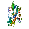
 Mass: 18.015 Da / Num. of mol.: 30 / Source method: isolated from a natural source / Formula: H2O
 Sample preparation
 / Beamline: 19-ID / Wavelength: 0.9794 Å
 Processing