 Movie
Movie Controller

[English] 日本語
 Yorodumi
- PDB-6itx: Structure of the C-terminal head domain of the avian adenovirus E... -
+ Open data

- Basic information
| Entry | Database: PDB / ID: 6itx | ||||||
|---|---|---|---|---|---|---|---|
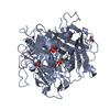
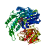
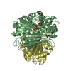
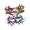
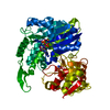
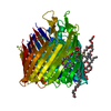
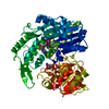
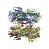
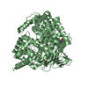
| Title | Structure of the C-terminal head domain of the avian adenovirus EDSV fiber | ||||||
 Components Components | Fiber protein | ||||||
 Keywords Keywords | CELL ADHESION / avian adenovirus / fiber protein | ||||||
| Function / homology |  Function and homology information Function and homology informationadhesion receptor-mediated virion attachment to host cell / virion component / cell adhesion / symbiont entry into host cell / host cell nucleus Similarity search - Function | ||||||
| Biological species |  Duck adenovirus 1 Duck adenovirus 1 | ||||||
| Method |  X-RAY DIFFRACTION / Resolution: 2.75 Å X-RAY DIFFRACTION / Resolution: 2.75 Å | ||||||
 Authors Authors | Wei, Q. / Song, Y. | ||||||
| Funding support |  China, 1items China, 1items
| ||||||
 Citation Citation | Journal: Int.J.Biol.Macromol. / Year: 2019 Title: Unravelling the receptor binding property of egg drop syndrome virus (EDSV) from the crystal structure of EDSV fiber head. Authors: Song, Y. / Wei, Q. / Liu, Y. / Feng, H. / Chen, Y. / Wang, Y. / Bai, Y. / Xing, G. / Deng, R. / Zhang, G. | ||||||
| History |
|
- Structure visualization
| Structure viewer | Molecule: MolmilJmol/JSmol |
|---|
- Downloads & links
-Download
| PDBx/mmCIF format | 6itx.cif.gz | 265.7 KB | Display | PDBx/mmCIF format |
|---|---|---|---|---|
| PDB format | pdb6itx.ent.gz | 211.9 KB | Display | PDB format |
| PDBx/mmJSON format | 6itx.json.gz | Tree view | PDBx/mmJSON format | |
| Others |  Other downloads Other downloads |
-Validation report
| Arichive directory | https://data.pdbj.org/pub/pdb/validation_reports/it/6itxftp://data.pdbj.org/pub/pdb/validation_reports/it/6itx | HTTPS FTP |
|---|
-Related structure data
| Similar structure data |
|---|
-Links
 PDBj
PDBj
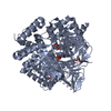
- Assembly
| Deposited unit | 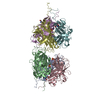
| ||||||||
|---|---|---|---|---|---|---|---|---|---|
| 1 | 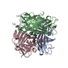
| ||||||||
| 2 | 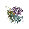
| ||||||||
| Unit cell |
|
-Components
| #1: Protein | Mass: 32100.943 Da / Num. of mol.: 6 Source method: isolated from a genetically manipulated source Source: (gene. exp.) Duck adenovirus 1 / Production host:  #2: Chemical | ChemComp-GLN /   Type: L-peptide linking / Mass: 146.144 Da / Num. of mol.: 5 / Source method: obtained synthetically / Formula: C5H10N2O3 Type: L-peptide linking / Mass: 146.144 Da / Num. of mol.: 5 / Source method: obtained synthetically / Formula: C5H10N2O3#3: Chemical | ChemComp-SO4 /   Mass: 96.063 Da / Num. of mol.: 5 / Source method: obtained synthetically / Formula: SO4 Mass: 96.063 Da / Num. of mol.: 5 / Source method: obtained synthetically / Formula: SO4#4: Water | ChemComp-HOH / |  Mass: 18.015 Da / Num. of mol.: 255 / Source method: isolated from a natural source / Formula: H2O Mass: 18.015 Da / Num. of mol.: 255 / Source method: isolated from a natural source / Formula: H2O |
|---|
-Experimental details
-Experiment
| Experiment | Method: X-RAY DIFFRACTION / Number of used crystals: 1 |
|---|
- Sample preparation
| Crystal | Density Matthews: 2.86 Å3/Da / Density % sol: 56.92 % |
|---|---|
| Crystal grow | Temperature: 293.15 K / Method: evaporation / Details: 0.2M Potassium sulfate, 15% w/v PEG 3350 |
-Data collection
| Diffraction | Mean temperature: 285 K / Serial crystal experiment: N | |||||||||||||||||||||||||||||||||||||||||||||
|---|---|---|---|---|---|---|---|---|---|---|---|---|---|---|---|---|---|---|---|---|---|---|---|---|---|---|---|---|---|---|---|---|---|---|---|---|---|---|---|---|---|---|---|---|---|---|
| Diffraction source | Source: ROTATING ANODE / Type: ENRAF-NONIUS FR591 / Wavelength: 0.987 Å | |||||||||||||||||||||||||||||||||||||||||||||
| Detector | Type: ADSC QUANTUM 210r / Detector: CCD / Date: May 20, 2017 | |||||||||||||||||||||||||||||||||||||||||||||
| Radiation | Protocol: SINGLE WAVELENGTH / Monochromatic (M) / Laue (L): M / Scattering type: x-ray | |||||||||||||||||||||||||||||||||||||||||||||
| Radiation wavelength | Wavelength: 0.987 Å / Relative weight: 1 | |||||||||||||||||||||||||||||||||||||||||||||
| Reflection | Resolution: 2.75→30 Å / Num. obs: 57643 / % possible obs: 99.8 % / Redundancy: 6.5 % / Biso Wilson estimate: 46.9 Å2 / Rmerge(I) obs: 0.121 / Rpim(I) all: 0.051 / Rrim(I) all: 0.131 / Χ2: 0.99 / Net I/σ(I): 5.6 | |||||||||||||||||||||||||||||||||||||||||||||
| Reflection shell | Diffraction-ID: 1
|
- Processing
| Software |
| ||||||||||||||||||||||||||||||||||||||||||||||||||||||||||||||||||||||||||||||||||||||||||||||||||||||||||||||||||||||||||||||||||||||||||||||||||||||||||
|---|---|---|---|---|---|---|---|---|---|---|---|---|---|---|---|---|---|---|---|---|---|---|---|---|---|---|---|---|---|---|---|---|---|---|---|---|---|---|---|---|---|---|---|---|---|---|---|---|---|---|---|---|---|---|---|---|---|---|---|---|---|---|---|---|---|---|---|---|---|---|---|---|---|---|---|---|---|---|---|---|---|---|---|---|---|---|---|---|---|---|---|---|---|---|---|---|---|---|---|---|---|---|---|---|---|---|---|---|---|---|---|---|---|---|---|---|---|---|---|---|---|---|---|---|---|---|---|---|---|---|---|---|---|---|---|---|---|---|---|---|---|---|---|---|---|---|---|---|---|---|---|---|---|---|---|
| Refinement | Resolution: 2.75→29.882 Å / SU ML: 0.29 / Cross valid method: THROUGHOUT / σ(F): 1.35 / Phase error: 24.28
| ||||||||||||||||||||||||||||||||||||||||||||||||||||||||||||||||||||||||||||||||||||||||||||||||||||||||||||||||||||||||||||||||||||||||||||||||||||||||||
| Solvent computation | Shrinkage radii: 0.9 Å / VDW probe radii: 1.11 Å | ||||||||||||||||||||||||||||||||||||||||||||||||||||||||||||||||||||||||||||||||||||||||||||||||||||||||||||||||||||||||||||||||||||||||||||||||||||||||||
| Displacement parameters | Biso max: 122.8 Å2 / Biso mean: 46.6149 Å2 / Biso min: 13.64 Å2 | ||||||||||||||||||||||||||||||||||||||||||||||||||||||||||||||||||||||||||||||||||||||||||||||||||||||||||||||||||||||||||||||||||||||||||||||||||||||||||
| Refinement step | Cycle: final / Resolution: 2.75→29.882 Å
| ||||||||||||||||||||||||||||||||||||||||||||||||||||||||||||||||||||||||||||||||||||||||||||||||||||||||||||||||||||||||||||||||||||||||||||||||||||||||||
| Refine LS restraints |
| ||||||||||||||||||||||||||||||||||||||||||||||||||||||||||||||||||||||||||||||||||||||||||||||||||||||||||||||||||||||||||||||||||||||||||||||||||||||||||
| LS refinement shell | Refine-ID: X-RAY DIFFRACTION / Rfactor Rfree error: 0 / Total num. of bins used: 21
|
