 Movie
Movie Controller

[English] 日本語
 Yorodumi
- PDB-6itk: Crystal structure of malate dehydrogenase from Corynebacterium gl... -
+ Open data

- Basic information
| Entry | Database: PDB / ID: 6itk | ||||||
|---|---|---|---|---|---|---|---|
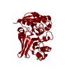
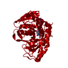
| Title | Crystal structure of malate dehydrogenase from Corynebacterium glutamicum ATCC 13032 in complex with NAD and malate | ||||||
 Components Components | Malate dehydrogenase | ||||||
 Keywords Keywords | OXIDOREDUCTASE / dehydrogenase | ||||||
| Function / homology |  Function and homology information Function and homology information(S)-malate dehydrogenase (NAD+, oxaloacetate-forming) / L-malate dehydrogenase (NAD+) activity / malate metabolic process / tricarboxylic acid cycle Similarity search - Function | ||||||
| Biological species |  Corynebacterium glutamicum ATCC 13032 (bacteria) Corynebacterium glutamicum ATCC 13032 (bacteria) | ||||||
| Method |  X-RAY DIFFRACTION / SYNCHROTRON / MOLECULAR REPLACEMENT / Resolution: 2 Å X-RAY DIFFRACTION / SYNCHROTRON / MOLECULAR REPLACEMENT / Resolution: 2 Å | ||||||
 Authors Authors | Seo, H. / Kim, K.-J. | ||||||
 Citation Citation | Journal: Nat Commun / Year: 2020 Title: Enhanced succinic acid production by Mannheimia employing optimal malate dehydrogenase. Authors: Ahn, J.H. / Seo, H. / Park, W. / Seok, J. / Lee, J.A. / Kim, W.J. / Kim, G.B. / Kim, K.J. / Lee, S.Y. | ||||||
| History |
|
- Structure visualization
| Structure viewer | Molecule: MolmilJmol/JSmol |
|---|
- Downloads & links
-Download
| PDBx/mmCIF format | 6itk.cif.gz | 138.3 KB | Display | PDBx/mmCIF format |
|---|---|---|---|---|
| PDB format | pdb6itk.ent.gz | 106.7 KB | Display | PDB format |
| PDBx/mmJSON format | 6itk.json.gz | Tree view | PDBx/mmJSON format | |
| Others |  Other downloads Other downloads |
-Validation report
| Arichive directory | https://data.pdbj.org/pub/pdb/validation_reports/it/6itkftp://data.pdbj.org/pub/pdb/validation_reports/it/6itk | HTTPS FTP |
|---|
-Related structure data
| Related structure data |  6itlC  4tvoS S: Starting model for refinement C: citing same article ( |
|---|---|
| Similar structure data |


-Links
 PDBj
PDBj


- Assembly
| Deposited unit | 
| ||||||||
|---|---|---|---|---|---|---|---|---|---|
| 1 |
| ||||||||
| Unit cell |
| ||||||||
| Components on special symmetry positions |
|
-Components
| #1: Protein | Mass: 36011.082 Da / Num. of mol.: 2 Source method: isolated from a genetically manipulated source Source: (gene. exp.) Corynebacterium glutamicum ATCC 13032 (bacteria)Strain: ATCC 13032 / DSM 20300 / JCM 1318 / LMG 3730 / NCIMB 10025 Gene: mdh, Cgl2380, cg2613 / Plasmid: pET30a / Production host: References: UniProt: Q8NN33, (S)-malate dehydrogenase (NAD+, oxaloacetate-forming) #2: Chemical |   Mass: 134.087 Da / Num. of mol.: 2 / Source method: obtained synthetically / Formula: C4H6O5 Mass: 134.087 Da / Num. of mol.: 2 / Source method: obtained synthetically / Formula: C4H6O5#3: Chemical |   Mass: 663.425 Da / Num. of mol.: 2 / Source method: obtained synthetically / Formula: C21H27N7O14P2 / Comment: NAD*YM Mass: 663.425 Da / Num. of mol.: 2 / Source method: obtained synthetically / Formula: C21H27N7O14P2 / Comment: NAD*YM#4: Water | ChemComp-HOH / |  Mass: 18.015 Da / Num. of mol.: 229 / Source method: isolated from a natural source / Formula: H2O Mass: 18.015 Da / Num. of mol.: 229 / Source method: isolated from a natural source / Formula: H2OHas ligand of interest | N | |
|---|
-Experimental details
-Experiment
| Experiment | Method: X-RAY DIFFRACTION / Number of used crystals: 1 |
|---|
- Sample preparation
| Crystal | Density Matthews: 2.75 Å3/Da / Density % sol: 55.21 % |
|---|---|
| Crystal grow | Temperature: 293 K / Method: vapor diffusion, hanging drop / pH: 7.5 / Details: PEG 3350, HEPES, MgCl2 |
-Data collection
| Diffraction | Mean temperature: 100 K / Serial crystal experiment: N |
|---|---|
| Diffraction source | Source: SYNCHROTRON / Site: PAL/PLS  / Beamline: 7A (6B, 6C1) / Wavelength: 0.97934 Å / Beamline: 7A (6B, 6C1) / Wavelength: 0.97934 Å |
| Detector | Type: ADSC QUANTUM 270 / Detector: CCD / Date: Dec 18, 2016 |
| Radiation | Monochromator: Double Crystal Monochromator / Protocol: SINGLE WAVELENGTH / Monochromatic (M) / Laue (L): M / Scattering type: x-ray |
| Radiation wavelength | Wavelength: 0.97934 Å / Relative weight: 1 |
| Reflection | Resolution: 2→77.08 Å / Num. obs: 50050 / % possible obs: 96.3 % / Redundancy: 2.7 % / Rmerge(I) obs: 0.123 / Net I/σ(I): 18 |
| Reflection shell | Resolution: 2→2.03 Å / Rmerge(I) obs: 0.39 / Num. unique obs: 2392 / CC1/2: 0.792 / % possible all: 93 |
- Processing
| Software |
| ||||||||||||||||||||||||||||||||||||||||||||||||||||||||||||
|---|---|---|---|---|---|---|---|---|---|---|---|---|---|---|---|---|---|---|---|---|---|---|---|---|---|---|---|---|---|---|---|---|---|---|---|---|---|---|---|---|---|---|---|---|---|---|---|---|---|---|---|---|---|---|---|---|---|---|---|---|---|
| Refinement | Method to determine structure: MOLECULAR REPLACEMENT Starting model: 4TVO Resolution: 2→26.73 Å / Cor.coef. Fo:Fc: 0.948 / Cor.coef. Fo:Fc free: 0.925 / SU B: 4.883 / SU ML: 0.128 / Cross valid method: THROUGHOUT / σ(F): 0 / ESU R: 0.17 / ESU R Free: 0.155 / Stereochemistry target values: MAXIMUM LIKELIHOOD Details: HYDROGENS HAVE BEEN ADDED IN THE RIDING POSITIONS U VALUES : REFINED INDIVIDUALLY
| ||||||||||||||||||||||||||||||||||||||||||||||||||||||||||||
| Solvent computation | Ion probe radii: 0.8 Å / Shrinkage radii: 0.8 Å / VDW probe radii: 1.2 Å / Solvent model: MASK | ||||||||||||||||||||||||||||||||||||||||||||||||||||||||||||
| Displacement parameters | Biso max: 89.39 Å2 / Biso mean: 24.369 Å2 / Biso min: 12.24 Å2
| ||||||||||||||||||||||||||||||||||||||||||||||||||||||||||||
| Refinement step | Cycle: final / Resolution: 2→26.73 Å
| ||||||||||||||||||||||||||||||||||||||||||||||||||||||||||||
| Refine LS restraints |
| ||||||||||||||||||||||||||||||||||||||||||||||||||||||||||||
| LS refinement shell | Resolution: 2.003→2.055 Å / Rfactor Rfree error: 0 / Total num. of bins used: 20
|
