 Movie
Movie Controller


+ Open data

- Basic information
| Entry | Database: PDB / ID: 6isk | ||||||
|---|---|---|---|---|---|---|---|
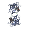
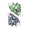
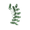
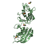
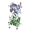
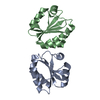
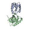
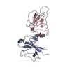
| Title | SnoaL-like cyclase XimE | ||||||
 Components Components | XimE, SnoaL-like domain protein | ||||||
 Keywords Keywords | LYASE / Snoal-like cyclase | ||||||
| Function / homology | SnoaL-like domain / SnoaL-like domain / NTF2-like domain superfamily / MALONIC ACID / XimE Function and homology information Function and homology information | ||||||
| Biological species |  Streptomyces xiamenensis 318 (bacteria) Streptomyces xiamenensis 318 (bacteria) | ||||||
| Method |  X-RAY DIFFRACTION / SYNCHROTRON / SAD / Resolution: 1.77 Å X-RAY DIFFRACTION / SYNCHROTRON / SAD / Resolution: 1.77 Å | ||||||
 Authors Authors | He, B. / Zhou, T. / Bu, X. / Weng, J. / Xu, J. / Lin, S. / Zheng, J. / Zhao, Y. / Xu, M. | ||||||
| Funding support |  China, 1items China, 1items
| ||||||
 Citation Citation | Journal: Acs Catalysis / Year: 2019 Title: Enzymatic Pyran Formation Involved in Xiamenmycin Biosynthesis Authors: He, B. / Zhou, T. / Bu, X. / Weng, J. / Xu, J. / Lin, S. / Zheng, J. / Zhao, Y. / Xu, M. | ||||||
| History |
|
- Structure visualization
| Structure viewer | Molecule: MolmilJmol/JSmol |
|---|
- Downloads & links
-Download
| PDBx/mmCIF format | 6isk.cif.gz | 62 KB | Display | PDBx/mmCIF format |
|---|---|---|---|---|
| PDB format | pdb6isk.ent.gz | 44.7 KB | Display | PDB format |
| PDBx/mmJSON format | 6isk.json.gz | Tree view | PDBx/mmJSON format | |
| Others |  Other downloads Other downloads |
-Validation report
| Arichive directory | https://data.pdbj.org/pub/pdb/validation_reports/is/6iskftp://data.pdbj.org/pub/pdb/validation_reports/is/6isk | HTTPS FTP |
|---|
-Related structure data


-Links
 PDBj
PDBj

- Assembly
| Deposited unit | 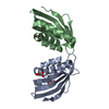
| ||||||||
|---|---|---|---|---|---|---|---|---|---|
| 1 | 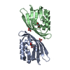
| ||||||||
| Unit cell |
|
-Components
| #1: Protein | Mass: 17205.289 Da / Num. of mol.: 2 Source method: isolated from a genetically manipulated source Source: (gene. exp.) Streptomyces xiamenensis 318 (bacteria)Gene: ximE / Plasmid: pET28a(+) / Production host: References: UniProt: U5Q2N8, Lyases; Carbon-oxygen lyases; Other carbon-oxygen lyases #2: Chemical |   Mass: 104.061 Da / Num. of mol.: 3 Mass: 104.061 Da / Num. of mol.: 3Source method: isolated from a genetically manipulated source Formula: C3H4O4 / Feature type: SUBJECT OF INVESTIGATION #3: Chemical | ChemComp-EDO / |   Mass: 62.068 Da / Num. of mol.: 1 / Source method: obtained synthetically / Formula: C2H6O2 / Feature type: SUBJECT OF INVESTIGATION Mass: 62.068 Da / Num. of mol.: 1 / Source method: obtained synthetically / Formula: C2H6O2 / Feature type: SUBJECT OF INVESTIGATION#4: Water | ChemComp-HOH / |  Mass: 18.015 Da / Num. of mol.: 52 / Source method: isolated from a natural source / Formula: H2O Mass: 18.015 Da / Num. of mol.: 52 / Source method: isolated from a natural source / Formula: H2O |
|---|
-Experimental details
-Experiment
| Experiment | Method: X-RAY DIFFRACTION / Number of used crystals: 1 |
|---|
- Sample preparation
| Crystal | Density Matthews: 1.99 Å3/Da / Density % sol: 38.32 % / Description: Starked layers of thin slices. |
|---|---|
| Crystal grow | Temperature: 293 K / Method: vapor diffusion, sitting drop / pH: 4 / Details: 0.2M malonic acid, 22% PEG 3350, pH 4.0 / PH range: 3.8-4.5 / Temp details: 293 |
-Data collection
| Diffraction | Mean temperature: 100 K / Ambient temp details: 100 / Serial crystal experiment: N |
|---|---|
| Diffraction source | Source: SYNCHROTRON / Site: SSRF / Beamline: BL18U1 / Wavelength: 0.9778 Å |
| Detector | Type: DECTRIS PILATUS3 6M / Detector: PIXEL / Date: Nov 22, 2017 |
| Radiation | Protocol: SINGLE WAVELENGTH / Monochromatic (M) / Laue (L): M / Scattering type: x-ray |
| Radiation wavelength | Wavelength: 0.9778 Å / Relative weight: 1 |
| Reflection | Resolution: 1.77→59 Å / Num. obs: 25607 / % possible obs: 97.6 % / Redundancy: 3.7 % / Rmerge(I) obs: 0.088 / Net I/σ(I): 15.4 |
| Reflection shell | Resolution: 1.77→1.8 Å / Redundancy: 3.2 % / Rmerge(I) obs: 0.438 / % possible all: 83 |
- Processing
| Software |
| ||||||||||||||||||||||||||||||||||||||||||||||||||||||||||||
|---|---|---|---|---|---|---|---|---|---|---|---|---|---|---|---|---|---|---|---|---|---|---|---|---|---|---|---|---|---|---|---|---|---|---|---|---|---|---|---|---|---|---|---|---|---|---|---|---|---|---|---|---|---|---|---|---|---|---|---|---|---|
| Refinement | Method to determine structure: SAD / Resolution: 1.77→58.82 Å / Cor.coef. Fo:Fc: 0.935 / Cor.coef. Fo:Fc free: 0.912 / SU B: 2.763 / SU ML: 0.088 / Cross valid method: THROUGHOUT / σ(F): 0 / ESU R: 0.128 / ESU R Free: 0.122 Details: HYDROGENS HAVE BEEN ADDED IN THE RIDING POSITIONS U VALUES : REFINED INDIVIDUALLY
| ||||||||||||||||||||||||||||||||||||||||||||||||||||||||||||
| Solvent computation | Ion probe radii: 0.8 Å / Shrinkage radii: 0.8 Å / VDW probe radii: 1.2 Å | ||||||||||||||||||||||||||||||||||||||||||||||||||||||||||||
| Displacement parameters | Biso max: 74.7 Å2 / Biso mean: 23.098 Å2 / Biso min: 9.31 Å2
| ||||||||||||||||||||||||||||||||||||||||||||||||||||||||||||
| Refinement step | Cycle: final / Resolution: 1.77→58.82 Å
| ||||||||||||||||||||||||||||||||||||||||||||||||||||||||||||
| Refine LS restraints |
| ||||||||||||||||||||||||||||||||||||||||||||||||||||||||||||
| LS refinement shell | Resolution: 1.766→1.812 Å / Rfactor Rfree error: 0 / Total num. of bins used: 20
|
