 Movie
Movie Controller


+ Open data

- Basic information
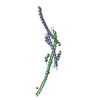
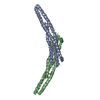
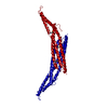
| Entry | Database: PDB / ID: 6ird | ||||||
|---|---|---|---|---|---|---|---|
| Title | Complex structure of INADL PDZ89 and PLCb4 C-terminal CC-PBM | ||||||
 Components Components |
| ||||||
 Keywords Keywords | HYDROLASE/PROTEIN BINDING / PDZ supramodule / phospholipase C beta / HYDROLASE-PROTEIN BINDING complex | ||||||
| Function / homology |  Function and homology information Function and homology informationPLC beta mediated events / Synthesis of IP3 and IP4 in the cytosol / tight junction assembly / Tight junction interactions / SARS-CoV-2 targets PDZ proteins in cell-cell junction / phosphoinositide phospholipase C / establishment of apical/basal cell polarity / G alpha (q) signalling events / phosphatidylinositol metabolic process / phosphatidylinositol-4,5-bisphosphate phospholipase C activity ...PLC beta mediated events / Synthesis of IP3 and IP4 in the cytosol / tight junction assembly / Tight junction interactions / SARS-CoV-2 targets PDZ proteins in cell-cell junction / phosphoinositide phospholipase C / establishment of apical/basal cell polarity / G alpha (q) signalling events / phosphatidylinositol metabolic process / phosphatidylinositol-4,5-bisphosphate phospholipase C activity / establishment or maintenance of epithelial cell apical/basal polarity / mitogen-activated protein kinase binding / apical junction complex / smooth endoplasmic reticulum / phosphatidylinositol-mediated signaling / negative regulation of potassium ion transport / parallel fiber to Purkinje cell synapse / bicellular tight junction / lipid catabolic process / release of sequestered calcium ion into cytosol / modulation of chemical synaptic transmission / centriolar satellite / apical part of cell / cell junction / apical plasma membrane / postsynaptic density / intracellular signal transduction / postsynapse / calcium ion binding / dendrite / perinuclear region of cytoplasm / glutamatergic synapse / extracellular exosome / nucleus / plasma membrane / cytoplasm / cytosol Similarity search - Function | ||||||
| Biological species |   Homo sapiens (human) Homo sapiens (human) | ||||||
| Method |  X-RAY DIFFRACTION / SYNCHROTRON / MOLECULAR REPLACEMENT / Resolution: 2.813 Å X-RAY DIFFRACTION / SYNCHROTRON / MOLECULAR REPLACEMENT / Resolution: 2.813 Å | ||||||
 Authors Authors | Ye, F. / Li, J. / Huang, Y. / Liu, W. / Zhang, M. | ||||||
 Citation Citation | Journal: Elife / Year: 2018 Title: An unexpected INAD PDZ tandem-mediated plc beta binding in Drosophila photo receptors. Authors: Ye, F. / Huang, Y. / Li, J. / Ma, Y. / Xie, C. / Liu, Z. / Deng, X. / Wan, J. / Xue, T. / Liu, W. / Zhang, M. | ||||||
| History |
|
- Structure visualization
| Structure viewer | Molecule: MolmilJmol/JSmol |
|---|
- Downloads & links
-Download
| PDBx/mmCIF format | 6ird.cif.gz | 98.1 KB | Display | PDBx/mmCIF format |
|---|---|---|---|---|
| PDB format | pdb6ird.ent.gz | 69.6 KB | Display | PDB format |
| PDBx/mmJSON format | 6ird.json.gz | Tree view | PDBx/mmJSON format | |
| Others |  Other downloads Other downloads |
-Validation report
| Arichive directory | https://data.pdbj.org/pub/pdb/validation_reports/ir/6irdftp://data.pdbj.org/pub/pdb/validation_reports/ir/6ird | HTTPS FTP |
|---|
-Related structure data
| Related structure data | 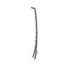 6irbC  6ircC  6ireC  3r0hS S: Starting model for refinement C: citing same article ( |
|---|---|
| Similar structure data |


-Links
 PDBj
PDBj


- Assembly
| Deposited unit | 
| ||||||||
|---|---|---|---|---|---|---|---|---|---|
| 1 |
| ||||||||
| Unit cell |
|
-Components
| #1: Protein | Mass: 31063.723 Da / Num. of mol.: 1 / Fragment: C-terminal CC-PBM / Mutation: K926A,K930A,K933A,K934A,K937A,K944A,K945A,K948A Source method: isolated from a genetically manipulated source Source: (gene. exp.)  References: UniProt: Q91UZ1, phosphoinositide phospholipase C |
|---|---|
| #2: Protein | Mass: 22723.523 Da / Num. of mol.: 1 / Fragment: PDZ89 Source method: isolated from a genetically manipulated source Source: (gene. exp.) Homo sapiens (human) / Gene: PATJ, INADL / Production host: |
| #3: Chemical | ChemComp-AU /   Mass: 196.967 Da / Num. of mol.: 5 / Source method: obtained synthetically / Formula: Au Mass: 196.967 Da / Num. of mol.: 5 / Source method: obtained synthetically / Formula: Au |
-Experimental details
-Experiment
| Experiment | Method: X-RAY DIFFRACTION / Number of used crystals: 1 |
|---|
- Sample preparation
| Crystal | Density Matthews: 2.61 Å3/Da / Density % sol: 52.86 % |
|---|---|
| Crystal grow | Temperature: 289 K / Method: liquid diffusion / pH: 6.5 Details: 0.2M MgCl2, 0.1M BIS-TRIS pH6.5, 20% w/v Polyethylene glycol 3350 |
-Data collection
| Diffraction | Mean temperature: 100 K / Serial crystal experiment: N | |||||||||||||||||||||||||||||||||||||||||||||||||||||||||||||||||||||||||||||||||||||||||||||||||||||||||||||||||||||||||||||||||||||||||||||||||||||||||||||||||||||||||||||||||||||||||||||
|---|---|---|---|---|---|---|---|---|---|---|---|---|---|---|---|---|---|---|---|---|---|---|---|---|---|---|---|---|---|---|---|---|---|---|---|---|---|---|---|---|---|---|---|---|---|---|---|---|---|---|---|---|---|---|---|---|---|---|---|---|---|---|---|---|---|---|---|---|---|---|---|---|---|---|---|---|---|---|---|---|---|---|---|---|---|---|---|---|---|---|---|---|---|---|---|---|---|---|---|---|---|---|---|---|---|---|---|---|---|---|---|---|---|---|---|---|---|---|---|---|---|---|---|---|---|---|---|---|---|---|---|---|---|---|---|---|---|---|---|---|---|---|---|---|---|---|---|---|---|---|---|---|---|---|---|---|---|---|---|---|---|---|---|---|---|---|---|---|---|---|---|---|---|---|---|---|---|---|---|---|---|---|---|---|---|---|---|---|---|---|
| Diffraction source | Source: SYNCHROTRON / Site: SSRF  / Beamline: BL17U1 / Wavelength: 0.97918 Å / Beamline: BL17U1 / Wavelength: 0.97918 Å | |||||||||||||||||||||||||||||||||||||||||||||||||||||||||||||||||||||||||||||||||||||||||||||||||||||||||||||||||||||||||||||||||||||||||||||||||||||||||||||||||||||||||||||||||||||||||||||
| Detector | Type: DECTRIS EIGER X 16M / Detector: PIXEL / Date: Mar 14, 2018 | |||||||||||||||||||||||||||||||||||||||||||||||||||||||||||||||||||||||||||||||||||||||||||||||||||||||||||||||||||||||||||||||||||||||||||||||||||||||||||||||||||||||||||||||||||||||||||||
| Radiation | Protocol: SINGLE WAVELENGTH / Monochromatic (M) / Laue (L): M / Scattering type: x-ray | |||||||||||||||||||||||||||||||||||||||||||||||||||||||||||||||||||||||||||||||||||||||||||||||||||||||||||||||||||||||||||||||||||||||||||||||||||||||||||||||||||||||||||||||||||||||||||||
| Radiation wavelength | Wavelength: 0.97918 Å / Relative weight: 1 | |||||||||||||||||||||||||||||||||||||||||||||||||||||||||||||||||||||||||||||||||||||||||||||||||||||||||||||||||||||||||||||||||||||||||||||||||||||||||||||||||||||||||||||||||||||||||||||
| Reflection | Resolution: 2.8→50 Å / Num. obs: 14070 / % possible obs: 99.7 % / Redundancy: 7.2 % / Rmerge(I) obs: 0.117 / Rpim(I) all: 0.048 / Rrim(I) all: 0.127 / Χ2: 1.676 / Net I/σ(I): 6.9 / Num. measured all: 100701 | |||||||||||||||||||||||||||||||||||||||||||||||||||||||||||||||||||||||||||||||||||||||||||||||||||||||||||||||||||||||||||||||||||||||||||||||||||||||||||||||||||||||||||||||||||||||||||||
| Reflection shell | Diffraction-ID: 1
|
- Processing
| Software |
| ||||||||||||||||||||||||
|---|---|---|---|---|---|---|---|---|---|---|---|---|---|---|---|---|---|---|---|---|---|---|---|---|---|
| Refinement | Method to determine structure: MOLECULAR REPLACEMENT Starting model: 3R0H Resolution: 2.813→14.997 Å / SU ML: 0.41 / Cross valid method: THROUGHOUT / σ(F): 0.31 / Phase error: 30.5 / Stereochemistry target values: ML
| ||||||||||||||||||||||||
| Solvent computation | Shrinkage radii: 0.9 Å / VDW probe radii: 1.11 Å / Solvent model: FLAT BULK SOLVENT MODEL | ||||||||||||||||||||||||
| Displacement parameters | Biso max: 300.44 Å2 / Biso mean: 28.8551 Å2 / Biso min: 2.09 Å2 | ||||||||||||||||||||||||
| Refinement step | Cycle: final / Resolution: 2.813→14.997 Å
| ||||||||||||||||||||||||
| LS refinement shell | Resolution: 2.8128→3.0936 Å
|
