 Movie
Movie Controller

[English] 日本語
 Yorodumi
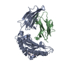
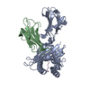
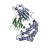
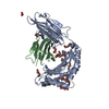
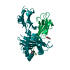
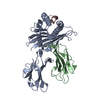
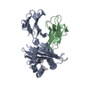
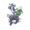
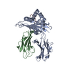
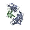
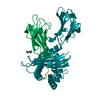
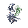
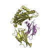
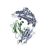
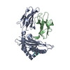
- PDB-6ilc: CRYSTAL STRUCTURE OF BAT MHC CLASS I PTAL-N*01:01 FOR 2.2 ANGSTROM -
+ Open data

- Basic information
| Entry | Database: PDB / ID: 6ilc | ||||||
|---|---|---|---|---|---|---|---|
| Title | CRYSTAL STRUCTURE OF BAT MHC CLASS I PTAL-N*01:01 FOR 2.2 ANGSTROM | ||||||
 Components Components |
| ||||||
 Keywords Keywords | IMMUNE SYSTEM / IMMUNOLOGY / VIRUS | ||||||
| Function / homology |  Function and homology information Function and homology informationresponse to metal ion / antigen processing and presentation of endogenous peptide antigen via MHC class Ib / antigen processing and presentation of endogenous peptide antigen via MHC class I via ER pathway, TAP-independent / antigen processing and presentation of peptide antigen via MHC class I / lumenal side of endoplasmic reticulum membrane / MHC class I protein complex / positive regulation of T cell mediated cytotoxicity / peptide antigen binding / phagocytic vesicle membrane / immune response ...response to metal ion / antigen processing and presentation of endogenous peptide antigen via MHC class Ib / antigen processing and presentation of endogenous peptide antigen via MHC class I via ER pathway, TAP-independent / antigen processing and presentation of peptide antigen via MHC class I / lumenal side of endoplasmic reticulum membrane / MHC class I protein complex / positive regulation of T cell mediated cytotoxicity / peptide antigen binding / phagocytic vesicle membrane / immune response / external side of plasma membrane / signaling receptor binding / : / extracellular region Similarity search - Function | ||||||
| Biological species |  Pteropus alecto (black flying fox) Pteropus alecto (black flying fox) Hendra virus Hendra virus | ||||||
| Method |  X-RAY DIFFRACTION / MOLECULAR REPLACEMENT / Resolution: 2.2 Å X-RAY DIFFRACTION / MOLECULAR REPLACEMENT / Resolution: 2.2 Å | ||||||
 Authors Authors | Qu, Z.H. / Zhang, N.Z. / Xia, C. | ||||||
 Citation Citation | Journal: J Immunol. / Year: 2019 Title: Structure and Peptidome of the Bat MHC Class I Molecule Reveal a Novel Mechanism Leading to High-Affinity Peptide Binding. Authors: Qu, Z. / Li, Z. / Ma, L. / Wei, X. / Zhang, L. / Liang, R. / Meng, G. / Zhang, N. / Xia, C. | ||||||
| History |
|
- Structure visualization
| Structure viewer | Molecule: MolmilJmol/JSmol |
|---|
- Downloads & links
-Download
| PDBx/mmCIF format | 6ilc.cif.gz | 177 KB | Display | PDBx/mmCIF format |
|---|---|---|---|---|
| PDB format | pdb6ilc.ent.gz | 140.8 KB | Display | PDB format |
| PDBx/mmJSON format | 6ilc.json.gz | Tree view | PDBx/mmJSON format | |
| Others |  Other downloads Other downloads |
-Validation report
| Arichive directory | https://data.pdbj.org/pub/pdb/validation_reports/il/6ilcftp://data.pdbj.org/pub/pdb/validation_reports/il/6ilc | HTTPS FTP |
|---|
-Related structure data
| Related structure data | 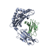 6ileC 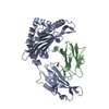 6ilfC 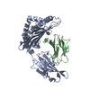 6ilgC 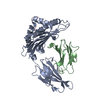 3vfnS S: Starting model for refinement C: citing same article ( |
|---|---|
| Similar structure data |


-Links
 PDBj
PDBj

- Assembly
| Deposited unit | 
| ||||||||
|---|---|---|---|---|---|---|---|---|---|
| 1 |
| ||||||||
| Unit cell |
| ||||||||
| Components on special symmetry positions |
|
-Components
| #1: Protein | Mass: 32345.482 Da / Num. of mol.: 1 / Fragment: UNP residues 25-303 Source method: isolated from a genetically manipulated source Source: (gene. exp.) Pteropus alecto (black flying fox) / Gene: Ptal-N / Production host:  |
|---|---|
| #2: Protein | Mass: 11474.836 Da / Num. of mol.: 1 Source method: isolated from a genetically manipulated source Source: (gene. exp.) Pteropus alecto (black flying fox) / Gene: PAL_GLEAN10023531 / Production host: |
| #3: Protein/peptide | Mass: 924.008 Da / Num. of mol.: 1 / Source method: obtained synthetically / Source: (synth.) Hendra virus |
| #4: Water | ChemComp-HOH /  Mass: 18.015 Da / Num. of mol.: 204 / Source method: isolated from a natural source / Formula: H2O Mass: 18.015 Da / Num. of mol.: 204 / Source method: isolated from a natural source / Formula: H2O |
| Has protein modification | Y |
| Sequence details | A NCBI Reference Sequence for Beta-2-microglobulin is XP_006920478.1. |
-Experimental details
-Experiment
| Experiment | Method: X-RAY DIFFRACTION / Number of used crystals: 1 |
|---|
- Sample preparation
| Crystal | Density Matthews: 2.39 Å3/Da / Density % sol: 48.57 % |
|---|---|
| Crystal grow | Temperature: 291 K / Method: vapor diffusion, hanging drop Details: 0.2M Lithium sulfate monohydrate, 0.1M BIS-TRIS ph6.5, 25%(w/v) Polyethylene glycol 3,350 |
-Data collection
| Diffraction | Mean temperature: 100 K / Serial crystal experiment: N |
|---|---|
| Diffraction source | Source: ROTATING ANODE / Type: RIGAKU MICROMAX-007 HF / Wavelength: 1.54178 Å |
| Detector | Type: RIGAKU RAXIS IV++ / Detector: IMAGE PLATE / Date: Oct 4, 2017 |
| Radiation | Monochromator: NI FILTER / Protocol: SINGLE WAVELENGTH / Monochromatic (M) / Laue (L): M / Scattering type: x-ray |
| Radiation wavelength | Wavelength: 1.54178 Å / Relative weight: 1 |
| Reflection | Resolution: 1.93→135.54 Å / Num. obs: 23392 / % possible obs: 99.8 % / Redundancy: 2 % / Rmerge(I) obs: 0.11 / Net I/σ(I): 16 |
| Reflection shell | Resolution: 1.93→1.98 Å / Rmerge(I) obs: 0.248 / Num. unique obs: 2127 |
- Processing
| Software |
| ||||||||||||||||||||||||||||||||||||||||||||||||||||||||||||||||||||||||||||||||||||||||||||||||||||
|---|---|---|---|---|---|---|---|---|---|---|---|---|---|---|---|---|---|---|---|---|---|---|---|---|---|---|---|---|---|---|---|---|---|---|---|---|---|---|---|---|---|---|---|---|---|---|---|---|---|---|---|---|---|---|---|---|---|---|---|---|---|---|---|---|---|---|---|---|---|---|---|---|---|---|---|---|---|---|---|---|---|---|---|---|---|---|---|---|---|---|---|---|---|---|---|---|---|---|---|---|---|
| Refinement | Method to determine structure: MOLECULAR REPLACEMENT Starting model: 3VFN Resolution: 2.2→14.9 Å / SU ML: 0.25 / Cross valid method: FREE R-VALUE / σ(F): 1.35 / Phase error: 23.57
| ||||||||||||||||||||||||||||||||||||||||||||||||||||||||||||||||||||||||||||||||||||||||||||||||||||
| Solvent computation | Shrinkage radii: 0.9 Å / VDW probe radii: 1.11 Å | ||||||||||||||||||||||||||||||||||||||||||||||||||||||||||||||||||||||||||||||||||||||||||||||||||||
| Refinement step | Cycle: LAST / Resolution: 2.2→14.9 Å
| ||||||||||||||||||||||||||||||||||||||||||||||||||||||||||||||||||||||||||||||||||||||||||||||||||||
| Refine LS restraints |
| ||||||||||||||||||||||||||||||||||||||||||||||||||||||||||||||||||||||||||||||||||||||||||||||||||||
| LS refinement shell |
| ||||||||||||||||||||||||||||||||||||||||||||||||||||||||||||||||||||||||||||||||||||||||||||||||||||
| Refinement TLS params. | Method: refined / Refine-ID: X-RAY DIFFRACTION
| ||||||||||||||||||||||||||||||||||||||||||||||||||||||||||||||||||||||||||||||||||||||||||||||||||||
| Refinement TLS group |
|
