 Movie
Movie Controller

[English] 日本語
 Yorodumi
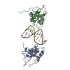
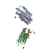
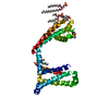
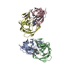
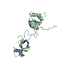
- PDB-6iir: Complex structure of the HRP3 PWWP domain with a 10-bp GC-rich DNA -
+ Open data

- Basic information
| Entry | Database: PDB / ID: 6iir | ||||||
|---|---|---|---|---|---|---|---|
| Title | Complex structure of the HRP3 PWWP domain with a 10-bp GC-rich DNA | ||||||
 Components Components |
| ||||||
 Keywords Keywords | DNA BINDING PROTEIN/DNA / growth factor / PWWP domain / Hepatoma-derived growth factor-related protein 3 / DNA BINDING PROTEIN / DNA BINDING PROTEIN-DNA complex | ||||||
| Function / homology |  Function and homology information Function and homology informationnegative regulation of microtubule depolymerization / microtubule polymerization / growth factor activity / tubulin binding / neuron projection development / microtubule binding / extracellular region / nucleoplasm / nucleus / cytosol Similarity search - Function | ||||||
| Biological species |  Homo sapiens (human) Homo sapiens (human)synthetic construct (others) | ||||||
| Method |  X-RAY DIFFRACTION / SYNCHROTRON / MOLECULAR REPLACEMENT / Resolution: 2.2 Å X-RAY DIFFRACTION / SYNCHROTRON / MOLECULAR REPLACEMENT / Resolution: 2.2 Å | ||||||
 Authors Authors | Wang, Z. / Tian, W. | ||||||
| Funding support |  China, 1items China, 1items
| ||||||
 Citation Citation | Journal: Nucleic Acids Res. / Year: 2019 Title: Complex structure of the HRP3 PWWP domain with a 10-bp GC-rich DNA Authors: Wang, Z. / Tian, W. | ||||||
| History |
|
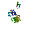
- Structure visualization
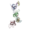
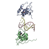
| Structure viewer | Molecule: MolmilJmol/JSmol |
|---|
- Downloads & links
-Download
| PDBx/mmCIF format | 6iir.cif.gz | 118.7 KB | Display | PDBx/mmCIF format |
|---|---|---|---|---|
| PDB format | pdb6iir.ent.gz | 91.8 KB | Display | PDB format |
| PDBx/mmJSON format | 6iir.json.gz | Tree view | PDBx/mmJSON format | |
| Others |  Other downloads Other downloads |
-Validation report
| Arichive directory | https://data.pdbj.org/pub/pdb/validation_reports/ii/6iirftp://data.pdbj.org/pub/pdb/validation_reports/ii/6iir | HTTPS FTP |
|---|
-Related structure data
| Related structure data | 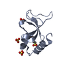 6iipS S: Starting model for refinement |
|---|---|
| Similar structure data |

-Links
 PDBj
PDBj
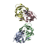
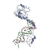
- Assembly
| Deposited unit | 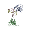
| ||||||||
|---|---|---|---|---|---|---|---|---|---|
| 1 |
| ||||||||
| Unit cell |
|
-Components
| #1: Protein | Mass: 11578.261 Da / Num. of mol.: 2 Source method: isolated from a genetically manipulated source Source: (gene. exp.) Homo sapiens (human) / Gene: HDGFL3, HDGF2, HDGFRP3, CGI-142 / Production host:  #2: DNA chain | Mass: 3046.981 Da / Num. of mol.: 2 / Source method: obtained synthetically / Source: (synth.) synthetic construct (others) #3: Water | ChemComp-HOH / |  Mass: 18.015 Da / Num. of mol.: 5 / Source method: isolated from a natural source / Formula: H2O Mass: 18.015 Da / Num. of mol.: 5 / Source method: isolated from a natural source / Formula: H2O |
|---|
-Experimental details
-Experiment
| Experiment | Method: X-RAY DIFFRACTION / Number of used crystals: 1 |
|---|
- Sample preparation
| Crystal | Density Matthews: 2.2 Å3/Da / Density % sol: 44.07 % |
|---|---|
| Crystal grow | Temperature: 293 K / Method: vapor diffusion, hanging drop / pH: 8 Details: 0.1 M Tris, pH 8.0, 0.1 M sodium malonate, pH 8.0, and 26% polyethylene glycol 1000 |
-Data collection
| Diffraction | Mean temperature: 77 K / Serial crystal experiment: N |
|---|---|
| Diffraction source | Source: SYNCHROTRON / Site: SSRF / Beamline: BL19U1 / Wavelength: 0.97853 Å |
| Detector | Type: DECTRIS PILATUS 6M / Detector: PIXEL / Date: Oct 26, 2016 |
| Radiation | Protocol: SINGLE WAVELENGTH / Monochromatic (M) / Laue (L): M / Scattering type: x-ray |
| Radiation wavelength | Wavelength: 0.97853 Å / Relative weight: 1 |
| Reflection | Resolution: 2.2→50 Å / Num. obs: 12832 / % possible obs: 99.9 % / Redundancy: 7.5 % / Rmerge(I) obs: 0.111 / Net I/σ(I): 35.9 |
| Reflection shell | Resolution: 2.2→2.24 Å / Redundancy: 7 % / Rmerge(I) obs: 0.975 / Mean I/σ(I) obs: 2.1 / Num. unique obs: 700 / % possible all: 100 |
- Processing
| Software |
| ||||||||||||||||||||||||||||||||||||||||
|---|---|---|---|---|---|---|---|---|---|---|---|---|---|---|---|---|---|---|---|---|---|---|---|---|---|---|---|---|---|---|---|---|---|---|---|---|---|---|---|---|---|
| Refinement | Method to determine structure: MOLECULAR REPLACEMENT Starting model: 6IIP Resolution: 2.2→29.059 Å / SU ML: 0.41 / Cross valid method: FREE R-VALUE / σ(F): 2.05 / Phase error: 37.72
| ||||||||||||||||||||||||||||||||||||||||
| Solvent computation | Shrinkage radii: 0.9 Å / VDW probe radii: 1.11 Å | ||||||||||||||||||||||||||||||||||||||||
| Refinement step | Cycle: LAST / Resolution: 2.2→29.059 Å
| ||||||||||||||||||||||||||||||||||||||||
| Refine LS restraints |
| ||||||||||||||||||||||||||||||||||||||||
| LS refinement shell |
| ||||||||||||||||||||||||||||||||||||||||
| Refinement TLS params. | Method: refined / Origin x: 56.163 Å / Origin y: -28.7894 Å / Origin z: -229.7076 Å
| ||||||||||||||||||||||||||||||||||||||||
| Refinement TLS group | Selection details: all |
