 Movie
Movie Controller

[English] 日本語
 Yorodumi
- PDB-6iey: Crystal structure of Chloramphenicol-Metabolizaing Enzyme EstDL13... -
+ Open data

- Basic information
| Entry | Database: PDB / ID: 6iey | ||||||||||||
|---|---|---|---|---|---|---|---|---|---|---|---|---|---|
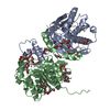
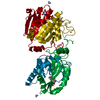
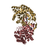
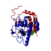
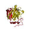
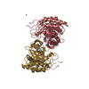
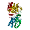
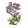
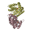
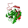
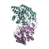
| Title | Crystal structure of Chloramphenicol-Metabolizaing Enzyme EstDL136-Chloramphenicol complex | ||||||||||||
 Components Components | Esterase | ||||||||||||
 Keywords Keywords | HYDROLASE / Chloramphenicol / metagenome / hornome sensitive lipase / HSL / EstDL136 / esterase | ||||||||||||
| Function / homology |  Function and homology information Function and homology information | ||||||||||||
| Biological species |  uncultured bacterium (environmental samples) uncultured bacterium (environmental samples) | ||||||||||||
| Method |  X-RAY DIFFRACTION / SYNCHROTRON / Resolution: 2.097 Å X-RAY DIFFRACTION / SYNCHROTRON / Resolution: 2.097 Å | ||||||||||||
 Authors Authors | Kim, S.H. / Kang, P.A. / Han, K.T. / Lee, S.W. / Rhee, S.K. | ||||||||||||
| Funding support |  Korea, Republic Of, 3items Korea, Republic Of, 3items
| ||||||||||||
 Citation Citation | Journal: PLoS ONE / Year: 2019 Title: Crystal structure of chloramphenicol-metabolizing enzyme EstDL136 from a metagenome. Authors: Kim, S.H. / Kang, P.A. / Han, K.T. / Lee, S.W. / Rhee, S.K. | ||||||||||||
| History |
|
- Structure visualization
| Structure viewer | Molecule: MolmilJmol/JSmol |
|---|
- Downloads & links
-Download
| PDBx/mmCIF format | 6iey.cif.gz | 131.8 KB | Display | PDBx/mmCIF format |
|---|---|---|---|---|
| PDB format | pdb6iey.ent.gz | 101.7 KB | Display | PDB format |
| PDBx/mmJSON format | 6iey.json.gz | Tree view | PDBx/mmJSON format | |
| Others |  Other downloads Other downloads |
-Validation report
| Arichive directory | https://data.pdbj.org/pub/pdb/validation_reports/ie/6ieyftp://data.pdbj.org/pub/pdb/validation_reports/ie/6iey | HTTPS FTP |
|---|
-Related structure data
-Links
 PDBj
PDBj- Assembly
| Deposited unit | 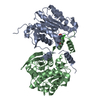
| ||||||||
|---|---|---|---|---|---|---|---|---|---|
| 1 |
| ||||||||
| Unit cell |
|
-Components
| #1: Protein | Mass: 34213.297 Da / Num. of mol.: 2 / Mutation: catalytic inactive mutant S156A Source method: isolated from a genetically manipulated source Details: To make EstDL136 crystal, internal residues from P37 to P39 were deleted Source: (gene. exp.) uncultured bacterium (environmental samples)Gene: estDL136 / Production host: #2: Chemical | ChemComp-CLM / |   Mass: 323.129 Da / Num. of mol.: 1 / Source method: isolated from a natural source / Formula: C11H12Cl2N2O5 / Comment: antibiotic*YM Mass: 323.129 Da / Num. of mol.: 1 / Source method: isolated from a natural source / Formula: C11H12Cl2N2O5 / Comment: antibiotic*YM#3: Water | ChemComp-HOH / |  Mass: 18.015 Da / Num. of mol.: 204 / Source method: isolated from a natural source / Formula: H2O Mass: 18.015 Da / Num. of mol.: 204 / Source method: isolated from a natural source / Formula: H2O |
|---|
-Experimental details
-Experiment
| Experiment | Method: X-RAY DIFFRACTION / Number of used crystals: 1 |
|---|
- Sample preparation
| Crystal | Density Matthews: 2.95 Å3/Da / Density % sol: 58.25 % |
|---|---|
| Crystal grow | Temperature: 295 K / Method: vapor diffusion, sitting drop / pH: 6.5 Details: 500mM Ammonium fluoride (pH6.5), 30% PEG 3350, 5% glyceol and 120mM TCEP. |
-Data collection
| Diffraction | Mean temperature: 100 K / Serial crystal experiment: N |
|---|---|
| Diffraction source | Source: SYNCHROTRON / Site: PAL/PLS / Beamline: 7A (6B, 6C1) / Wavelength: 0.97933 Å |
| Detector | Type: ADSC QUANTUM 270 / Detector: CCD / Date: Jun 26, 2014 |
| Radiation | Protocol: SINGLE WAVELENGTH / Monochromatic (M) / Laue (L): M / Scattering type: x-ray |
| Radiation wavelength | Wavelength: 0.97933 Å / Relative weight: 1 |
| Reflection | Resolution: 2.093→50 Å / Num. obs: 46963 / % possible obs: 96.4 % / Redundancy: 6.1 % / Net I/σ(I): 263 |
| Reflection shell | Resolution: 2.093→2.18 Å |
- Processing
| Software |
| |||||||||||||||||||||||||||||||||||||||||||||||||||||||||||||||||||||||||||||||||||||||||||||||||||||||||
|---|---|---|---|---|---|---|---|---|---|---|---|---|---|---|---|---|---|---|---|---|---|---|---|---|---|---|---|---|---|---|---|---|---|---|---|---|---|---|---|---|---|---|---|---|---|---|---|---|---|---|---|---|---|---|---|---|---|---|---|---|---|---|---|---|---|---|---|---|---|---|---|---|---|---|---|---|---|---|---|---|---|---|---|---|---|---|---|---|---|---|---|---|---|---|---|---|---|---|---|---|---|---|---|---|---|---|
| Refinement | Resolution: 2.097→29.04 Å / SU ML: 0.27 / Cross valid method: NONE / σ(F): 1.34 / Phase error: 25.16 / Stereochemistry target values: ML
| |||||||||||||||||||||||||||||||||||||||||||||||||||||||||||||||||||||||||||||||||||||||||||||||||||||||||
| Solvent computation | Shrinkage radii: 0.9 Å / VDW probe radii: 1.11 Å / Solvent model: FLAT BULK SOLVENT MODEL | |||||||||||||||||||||||||||||||||||||||||||||||||||||||||||||||||||||||||||||||||||||||||||||||||||||||||
| Refinement step | Cycle: LAST / Resolution: 2.097→29.04 Å
| |||||||||||||||||||||||||||||||||||||||||||||||||||||||||||||||||||||||||||||||||||||||||||||||||||||||||
| Refine LS restraints |
| |||||||||||||||||||||||||||||||||||||||||||||||||||||||||||||||||||||||||||||||||||||||||||||||||||||||||
| LS refinement shell |
|
