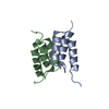
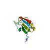
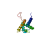
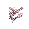
Evidence: gel filtration, In the previous gel filtration experiments, dNxf2-UBA co-migrated with Panx at a 1:1 ratio. In the construct for crystallization, we connected dNxf2-UBA (chain A) and Panx ...Evidence: gel filtration, In the previous gel filtration experiments, dNxf2-UBA co-migrated with Panx at a 1:1 ratio. In the construct for crystallization, we connected dNxf2-UBA (chain A) and Panx (chain B) using (Gly-Ser) linker.
Type
Name
Symmetry operation
Number
identity operation
1_555
x,y,z
1
Buried area
240 Å2
ΔGint
-1 kcal/mol
Surface area
6030 Å2
Method
PISA
Unit cell
Length a, b, c (Å)
29.074, 40.925, 83.091
Angle α, β, γ (deg.)
90.00, 90.00, 90.00
Int Tables number
18
Space group name H-M
P22121
-
Components
#1: Protein
FusionproteinofNuclearRNAexportfactor2andProteinpanoramix / Protein silencio
Mass: 10248.549 Da / Num. of mol.: 1 Source method: isolated from a genetically manipulated source Source: (gene. exp.) Drosophila melanogaster (fruit fly) / Gene: nxf2, CG4118, Panx, CG9754 / Production host: Escherichia coli (E. coli) / References: UniProt: Q9VV73, UniProt: Q9W2H9
In the structure databanks used in Yorodumi, some data are registered as the other names, "COVID-19 virus" and "2019-nCoV". Here are the details of the virus and the list of structure data.
Jan 31, 2019. EMDB accession codes are about to change! (news from PDBe EMDB page)
EMDB accession codes are about to change! (news from PDBe EMDB page)
The allocation of 4 digits for EMDB accession codes will soon come to an end. Whilst these codes will remain in use, new EMDB accession codes will include an additional digit and will expand incrementally as the available range of codes is exhausted. The current 4-digit format prefixed with “EMD-” (i.e. EMD-XXXX) will advance to a 5-digit format (i.e. EMD-XXXXX), and so on. It is currently estimated that the 4-digit codes will be depleted around Spring 2019, at which point the 5-digit format will come into force.
The EM Navigator/Yorodumi systems omit the EMD- prefix.
Related info.:Q: What is EMD? / ID/Accession-code notation in Yorodumi/EM Navigator
Yorodumi is a browser for structure data from EMDB, PDB, SASBDB, etc.
This page is also the successor to EM Navigator detail page, and also detail information page/front-end page for Omokage search.
The word "yorodu" (or yorozu) is an old Japanese word meaning "ten thousand". "mi" (miru) is to see.
Related info.:EMDB / PDB / SASBDB / Comparison of 3 databanks / Yorodumi Search / Aug 31, 2016. New EM Navigator & Yorodumi / Yorodumi Papers / Jmol/JSmol / Function and homology information / Changes in new EM Navigator and Yorodumi
 Movie
Movie Controller

 Yorodumi
 Open data
 Basic information
 Components
 Keywords
 Function and homology information
 X-RAY DIFFRACTION /
  Authors
 China, 2items
  Citation
 Structure visualization
 Downloads & links
 Other downloads
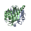


 PDBj
PDBj


 Assembly


 Mass: 92.094 Da / Num. of mol.: 1 / Source method: obtained synthetically / Formula: C3H8O3
 Mass: 18.015 Da / Num. of mol.: 216 / Source method: isolated from a natural source / Formula: H2O
 Sample preparation
 Processing