 Movie
Movie Controller


+ Open data

- Basic information
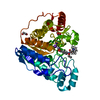
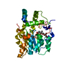
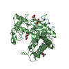
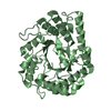
| Entry | Database: PDB / ID: 6idw | ||||||||||||
|---|---|---|---|---|---|---|---|---|---|---|---|---|---|
| Title | GH6 Orpinomyces sp. Y102 enzyme | ||||||||||||
 Components Components | Glucanase | ||||||||||||
 Keywords Keywords | HYDROLASE / Orpinomyces sp. / GH6 / Cbh7 / endoglucanases / exoglucanases | ||||||||||||
| Function / homology |  Function and homology information Function and homology informationHydrolases; Glycosylases; Glycosidases, i.e. enzymes that hydrolyse O- and S-glycosyl compounds / hydrolase activity, hydrolyzing O-glycosyl compounds / cellulose catabolic process Similarity search - Function | ||||||||||||
| Biological species |  Orpinomyces sp. Y102 (fungus) Orpinomyces sp. Y102 (fungus) | ||||||||||||
| Method |  X-RAY DIFFRACTION / SYNCHROTRON / MOLECULAR REPLACEMENT / Resolution: 2.78 Å X-RAY DIFFRACTION / SYNCHROTRON / MOLECULAR REPLACEMENT / Resolution: 2.78 Å | ||||||||||||
 Authors Authors | Tsai, L.C. / Huang, H.C. | ||||||||||||
| Funding support |  Taiwan, 1items Taiwan, 1items
| ||||||||||||
 Citation Citation | Journal: Acta Crystallogr D Struct Biol / Year: 2019 Title: Crystal structures of the GH6 Orpinomyces sp. Y102 CelC7 enzyme with exo and endo activity and its complex with cellobiose. Authors: Huang, H.C. / Qi, L.H. / Chen, Y.C. / Tsai, L.C. | ||||||||||||
| History |
|
- Structure visualization
| Structure viewer | Molecule: MolmilJmol/JSmol |
|---|
- Downloads & links
-Download
| PDBx/mmCIF format | 6idw.cif.gz | 263 KB | Display | PDBx/mmCIF format |
|---|---|---|---|---|
| PDB format | pdb6idw.ent.gz | 212.7 KB | Display | PDB format |
| PDBx/mmJSON format | 6idw.json.gz | Tree view | PDBx/mmJSON format | |
| Others |  Other downloads Other downloads |
-Validation report
| Arichive directory | https://data.pdbj.org/pub/pdb/validation_reports/id/6idwftp://data.pdbj.org/pub/pdb/validation_reports/id/6idw | HTTPS FTP |
|---|
-Related structure data
| Related structure data | 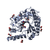 5jx5S S: Starting model for refinement |
|---|---|
| Similar structure data |


-Links
 PDBj
PDBj- Assembly
| Deposited unit | 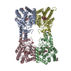
| ||||||||
|---|---|---|---|---|---|---|---|---|---|
| 1 | 
| ||||||||
| 2 | 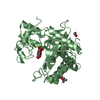
| ||||||||
| 3 | 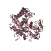
| ||||||||
| 4 | 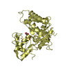
| ||||||||
| Unit cell |
| ||||||||
| Components on special symmetry positions |
|
-Components
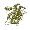
-Protein / Sugars , 2 types, 8 molecules ABCD
| #1: Protein | Mass: 35438.246 Da / Num. of mol.: 4 Source method: isolated from a genetically manipulated source Source: (gene. exp.) Orpinomyces sp. Y102 (fungus) / Gene: cbhC7Production host:  References: UniProt: A0A076U926, Hydrolases; Glycosylases; Glycosidases, i.e. enzymes that hydrolyse O- and S-glycosyl compounds #2: Polysaccharide | beta-D-glucopyranose-(1-4)-beta-D-glucopyranose / beta-cellobiose   Source method: isolated from a genetically manipulated source Details: oligosaccharide / References: beta-cellobiose |
|---|
-Non-polymers , 5 types, 360 molecules 


| #3: Chemical | ChemComp-GOL /  Mass: 92.094 Da / Num. of mol.: 4 / Source method: obtained synthetically / Formula: C3H8O3 Mass: 92.094 Da / Num. of mol.: 4 / Source method: obtained synthetically / Formula: C3H8O3#4: Chemical | ChemComp-PO4 / |  Mass: 94.971 Da / Num. of mol.: 1 / Source method: obtained synthetically / Formula: PO4 Mass: 94.971 Da / Num. of mol.: 1 / Source method: obtained synthetically / Formula: PO4#5: Chemical | ChemComp-EDO /  Mass: 62.068 Da / Num. of mol.: 4 / Source method: obtained synthetically / Formula: C2H6O2 Mass: 62.068 Da / Num. of mol.: 4 / Source method: obtained synthetically / Formula: C2H6O2#6: Chemical | ChemComp-MPD / ( |  Mass: 118.174 Da / Num. of mol.: 1 / Source method: obtained synthetically / Formula: C6H14O2 / Comment: precipitant*YM Mass: 118.174 Da / Num. of mol.: 1 / Source method: obtained synthetically / Formula: C6H14O2 / Comment: precipitant*YM#7: Water | ChemComp-HOH / | Mass: 18.015 Da / Num. of mol.: 350 / Source method: isolated from a natural source / Formula: H2O |
|---|
-Details
| Has protein modification | Y |
|---|
-Experimental details
-Experiment
| Experiment | Method: X-RAY DIFFRACTION / Number of used crystals: 1 |
|---|
- Sample preparation
| Crystal | Density Matthews: 3.89 Å3/Da / Density % sol: 68.41 % |
|---|---|
| Crystal grow | Temperature: 298 K / Method: vapor diffusion, hanging drop / pH: 7 / Details: 10% PEG 6K, 0.1M ammonium phosphate buffer, 2% MPD |
-Data collection
| Diffraction | Mean temperature: 100 K / Serial crystal experiment: N | |||||||||||||||||||||||||||||||||||||||||||||||||||||||||||||||||||||||||||||
|---|---|---|---|---|---|---|---|---|---|---|---|---|---|---|---|---|---|---|---|---|---|---|---|---|---|---|---|---|---|---|---|---|---|---|---|---|---|---|---|---|---|---|---|---|---|---|---|---|---|---|---|---|---|---|---|---|---|---|---|---|---|---|---|---|---|---|---|---|---|---|---|---|---|---|---|---|---|---|
| Diffraction source | Source: SYNCHROTRON / Site: NSRRC / Beamline: BL13B1 / Wavelength: 1 Å | |||||||||||||||||||||||||||||||||||||||||||||||||||||||||||||||||||||||||||||
| Detector | Type: ADSC QUANTUM 315r / Detector: CCD / Date: Mar 5, 2013 | |||||||||||||||||||||||||||||||||||||||||||||||||||||||||||||||||||||||||||||
| Radiation | Protocol: SINGLE WAVELENGTH / Monochromatic (M) / Laue (L): M / Scattering type: x-ray | |||||||||||||||||||||||||||||||||||||||||||||||||||||||||||||||||||||||||||||
| Radiation wavelength | Wavelength: 1 Å / Relative weight: 1 | |||||||||||||||||||||||||||||||||||||||||||||||||||||||||||||||||||||||||||||
| Reflection | Resolution: 2.78→154.87 Å / Num. obs: 52523 / % possible obs: 97.2 % / Redundancy: 4.3 % / Rmerge(I) obs: 0.055 / Χ2: 1.051 / Net I/σ(I): 29.6 | |||||||||||||||||||||||||||||||||||||||||||||||||||||||||||||||||||||||||||||
| Reflection shell |
|
- Processing
| Software |
| ||||||||||||||||||||||||
|---|---|---|---|---|---|---|---|---|---|---|---|---|---|---|---|---|---|---|---|---|---|---|---|---|---|
| Refinement | Method to determine structure: MOLECULAR REPLACEMENT Starting model: 5JX5 Resolution: 2.78→154.87 Å / Cor.coef. Fo:Fc: 0.888 / Cor.coef. Fo:Fc free: 0.848 / SU B: 0.002 / SU ML: 0 / Cross valid method: THROUGHOUT / σ(F): 0 / ESU R: 0.296 / ESU R Free: 0.34 / Details: U VALUES : REFINED INDIVIDUALLY
| ||||||||||||||||||||||||
| Solvent computation | Ion probe radii: 0.8 Å / Shrinkage radii: 0.8 Å / VDW probe radii: 1.2 Å | ||||||||||||||||||||||||
| Displacement parameters | Biso max: 90.34 Å2 / Biso mean: 32.426 Å2 / Biso min: 3.76 Å2
| ||||||||||||||||||||||||
| Refinement step | Cycle: final / Resolution: 2.78→154.87 Å
| ||||||||||||||||||||||||
| LS refinement shell | Resolution: 2.782→2.854 Å / Rfactor Rfree error: 0 / Total num. of bins used: 20
|
