| Entry | Database: PDB / ID: 6i9s
|
|---|
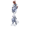
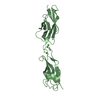
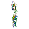
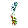
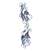
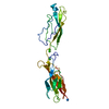
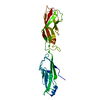
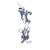
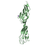
| Title | hRobo2 Extracellular Domains 2-3 |
|---|
 Components Components | Roundabout homolog 2 |
|---|
 Keywords Keywords | SIGNALING PROTEIN / Slit / Robo / Axon Guidance |
|---|
| Function / homology |  Function and homology information Function and homology information
olfactory bulb interneuron development / apoptotic process involved in luteolysis / negative regulation of negative chemotaxis / Regulation of cortical dendrite branching / negative regulation of synapse assembly / axon guidance receptor activity / heart induction / ROBO receptors bind AKAP5 / Formation of the ureteric bud / Regulation of commissural axon pathfinding by SLIT and ROBO ...olfactory bulb interneuron development / apoptotic process involved in luteolysis / negative regulation of negative chemotaxis / Regulation of cortical dendrite branching / negative regulation of synapse assembly / axon guidance receptor activity / heart induction / ROBO receptors bind AKAP5 / Formation of the ureteric bud / Regulation of commissural axon pathfinding by SLIT and ROBO / endocardial cushion formation / Signaling by ROBO receptors / pulmonary valve morphogenesis / metanephros development / outflow tract septum morphogenesis / axon midline choice point recognition / aortic valve morphogenesis / retinal ganglion cell axon guidance / ureteric bud development / positive regulation of axonogenesis / aorta development / ventricular septum morphogenesis / positive regulation of Notch signaling pathway / homophilic cell-cell adhesion / axolemma / cellular response to hormone stimulus / axon guidance / central nervous system development / cell-cell adhesion / brain development / chemotaxis / Regulation of expression of SLITs and ROBOs / cell surface / extracellular exosome / identical protein binding / plasma membraneSimilarity search - Function : / Immunoglobulin domain / Immunoglobulin I-set / Immunoglobulin I-set domain / Fibronectin type III domain / Fibronectin type 3 domain / Immunoglobulin subtype 2 / Immunoglobulin C-2 Type / Immunoglobulin V-Type / Fibronectin type-III domain profile. ...: / Immunoglobulin domain / Immunoglobulin I-set / Immunoglobulin I-set domain / Fibronectin type III domain / Fibronectin type 3 domain / Immunoglobulin subtype 2 / Immunoglobulin C-2 Type / Immunoglobulin V-Type / Fibronectin type-III domain profile. / Fibronectin type III / Fibronectin type III superfamily / Immunoglobulin V-set domain / Immunoglobulin subtype / Immunoglobulin / Ig-like domain profile. / Immunoglobulin-like domain / Immunoglobulin-like domain superfamily / Immunoglobulin-like fold / Immunoglobulins / Immunoglobulin-like / Sandwich / Mainly BetaSimilarity search - Domain/homology |
|---|
| Biological species |  Homo sapiens (human) Homo sapiens (human) |
|---|
| Method |  X-RAY DIFFRACTION / SYNCHROTRON / MOLECULAR REPLACEMENT / Resolution: 2.48 Å X-RAY DIFFRACTION / SYNCHROTRON / MOLECULAR REPLACEMENT / Resolution: 2.48 Å |
|---|
 Authors Authors | Barak, R. / Isupov, N.M. / Opatowsky, Y. |
|---|
| Funding support |  Israel, 1items Israel, 1items | Organization | Grant number | Country |
|---|
| Israel Science Foundation | 182/10 and 1425/15 | Israel |
|
|---|
 Citation Citation | Journal: Cell / Year: 2019
Title: Structural Principles in Robo Activation and Auto-inhibition.
Authors: Barak, R. / Yom-Tov, G. / Guez-Haddad, J. / Gasri-Plotnitsky, L. / Maimon, R. / Cohen-Berkman, M. / McCarthy, A.A. / Perlson, E. / Henis-Korenblit, S. / Isupov, M.N. / Opatowsky, Y. |
|---|
| History | | Deposition | Nov 25, 2018 | Deposition site: PDBE / Processing site: PDBE |
|---|
| Revision 1.0 | Mar 13, 2019 | Provider: repository / Type: Initial release |
|---|
| Revision 1.1 | Mar 20, 2019 | Group: Data collection / Database references / Category: citation / citation_author / pdbx_database_proc
Item: _citation.journal_abbrev / _citation.journal_id_CSD ..._citation.journal_abbrev / _citation.journal_id_CSD / _citation.journal_id_ISSN / _citation.pdbx_database_id_DOI / _citation.pdbx_database_id_PubMed / _citation.title / _citation.year |
|---|
| Revision 1.2 | Apr 17, 2019 | Group: Data collection / Database references
Category: citation / database_PDB_rev / database_PDB_rev_record
Item: _citation.journal_volume / _citation.page_first / _citation.page_last |
|---|
| Revision 1.3 | Nov 13, 2024 | Group: Data collection / Database references ...Data collection / Database references / Derived calculations / Structure summary
Category: chem_comp_atom / chem_comp_bond ...chem_comp_atom / chem_comp_bond / database_2 / pdbx_entry_details / pdbx_modification_feature / struct_conn
Item: _database_2.pdbx_DOI / _database_2.pdbx_database_accession ..._database_2.pdbx_DOI / _database_2.pdbx_database_accession / _struct_conn.pdbx_dist_value / _struct_conn.ptnr1_auth_asym_id / _struct_conn.ptnr1_auth_comp_id / _struct_conn.ptnr1_auth_seq_id / _struct_conn.ptnr1_label_asym_id / _struct_conn.ptnr1_label_atom_id / _struct_conn.ptnr1_label_comp_id / _struct_conn.ptnr1_label_seq_id / _struct_conn.ptnr2_auth_asym_id / _struct_conn.ptnr2_auth_comp_id / _struct_conn.ptnr2_auth_seq_id / _struct_conn.ptnr2_label_asym_id / _struct_conn.ptnr2_label_atom_id / _struct_conn.ptnr2_label_comp_id |
|---|
|
|---|
 Movie
Movie Controller


 Open data
 Basic information
 Structure visualization
 Downloads & links
 Other downloads


 PDBj
PDBj


 Assembly
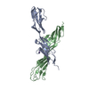

 Spodoptera frugiperda (fall armyworm) / References: UniProt: Q9HCK4
 Mass: 22.990 Da / Num. of mol.: 2 / Source method: obtained synthetically / Formula: Na
 Mass: 18.015 Da / Num. of mol.: 68 / Source method: isolated from a natural source / Formula: H2O
 Sample preparation
 / Beamline: 14.2 / Wavelength: 0.91 Å
 Processing