 Movie
Movie Controller

[English] 日本語
 Yorodumi
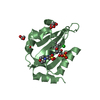
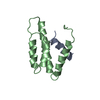
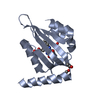
- PDB-6i22: Flavin Analogue Sheds Light on Light-Oxygen-Voltage Domain Mechanism -
+ Open data

- Basic information
| Entry | Database: PDB / ID: 6i22 | ||||||
|---|---|---|---|---|---|---|---|
| Title | Flavin Analogue Sheds Light on Light-Oxygen-Voltage Domain Mechanism | ||||||
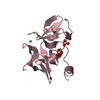
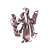
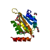
 Components Components | Aureochrome1-like protein | ||||||
 Keywords Keywords | TRANSCRIPTION / LOV Domain / FMN / Dark grown / fluorescence / light sensing / transcription factor PAS domain / Ochronomas danica | ||||||
| Function / homology |  Function and homology information Function and homology informationDNA-binding transcription factor activity / nucleotide binding / metal ion binding / nucleus Similarity search - Function | ||||||
| Biological species |  Ochromonas danica (eukaryote) Ochromonas danica (eukaryote) | ||||||
| Method |  X-RAY DIFFRACTION / SYNCHROTRON / MOLECULAR REPLACEMENT / Resolution: 1.66 Å X-RAY DIFFRACTION / SYNCHROTRON / MOLECULAR REPLACEMENT / Resolution: 1.66 Å | ||||||
 Authors Authors | Rizkallah, P.J. / Kalvaitis, M.E. / Allemann, R.K. / Mart, R.J. / Johnson, L.A. | ||||||
 Citation Citation | Journal: Biochemistry / Year: 2019 Title: A Noncanonical Chromophore Reveals Structural Rearrangements of the Light-Oxygen-Voltage Domain upon Photoactivation. Authors: Kalvaitis, M.E. / Johnson, L.A. / Mart, R.J. / Rizkallah, P. / Allemann, R.K. | ||||||
| History |
|
- Structure visualization
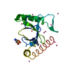
| Structure viewer | Molecule: MolmilJmol/JSmol |
|---|
- Downloads & links
-Download
| PDBx/mmCIF format | 6i22.cif.gz | 237.7 KB | Display | PDBx/mmCIF format |
|---|---|---|---|---|
| PDB format | pdb6i22.ent.gz | 191.9 KB | Display | PDB format |
| PDBx/mmJSON format | 6i22.json.gz | Tree view | PDBx/mmJSON format | |
| Others |  Other downloads Other downloads |
-Validation report
| Arichive directory | https://data.pdbj.org/pub/pdb/validation_reports/i2/6i22ftp://data.pdbj.org/pub/pdb/validation_reports/i2/6i22 | HTTPS FTP |
|---|
-Related structure data
| Related structure data | 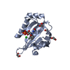 6i20C  6i21C  6i23C  6i24C  6i25C  5a8bS S: Starting model for refinement C: citing same article ( |
|---|---|
| Similar structure data |


-Links
 PDBj
PDBj


- Assembly
| Deposited unit | 
| ||||||||||||||||||||||||||||||||||||||||||||||||||||||||||||||||||||||||||||||||||||||||||||||||||||||||||||||||||||||||||||||||||||||||||||||||||||||||||||||||||||||||||||||||
|---|---|---|---|---|---|---|---|---|---|---|---|---|---|---|---|---|---|---|---|---|---|---|---|---|---|---|---|---|---|---|---|---|---|---|---|---|---|---|---|---|---|---|---|---|---|---|---|---|---|---|---|---|---|---|---|---|---|---|---|---|---|---|---|---|---|---|---|---|---|---|---|---|---|---|---|---|---|---|---|---|---|---|---|---|---|---|---|---|---|---|---|---|---|---|---|---|---|---|---|---|---|---|---|---|---|---|---|---|---|---|---|---|---|---|---|---|---|---|---|---|---|---|---|---|---|---|---|---|---|---|---|---|---|---|---|---|---|---|---|---|---|---|---|---|---|---|---|---|---|---|---|---|---|---|---|---|---|---|---|---|---|---|---|---|---|---|---|---|---|---|---|---|---|---|---|---|---|
| 1 | 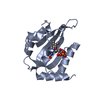
| ||||||||||||||||||||||||||||||||||||||||||||||||||||||||||||||||||||||||||||||||||||||||||||||||||||||||||||||||||||||||||||||||||||||||||||||||||||||||||||||||||||||||||||||||
| 2 | 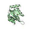
| ||||||||||||||||||||||||||||||||||||||||||||||||||||||||||||||||||||||||||||||||||||||||||||||||||||||||||||||||||||||||||||||||||||||||||||||||||||||||||||||||||||||||||||||||
| 3 | 
| ||||||||||||||||||||||||||||||||||||||||||||||||||||||||||||||||||||||||||||||||||||||||||||||||||||||||||||||||||||||||||||||||||||||||||||||||||||||||||||||||||||||||||||||||
| 4 | 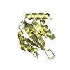
| ||||||||||||||||||||||||||||||||||||||||||||||||||||||||||||||||||||||||||||||||||||||||||||||||||||||||||||||||||||||||||||||||||||||||||||||||||||||||||||||||||||||||||||||||
| Unit cell |
| ||||||||||||||||||||||||||||||||||||||||||||||||||||||||||||||||||||||||||||||||||||||||||||||||||||||||||||||||||||||||||||||||||||||||||||||||||||||||||||||||||||||||||||||||
| Components on special symmetry positions |
| ||||||||||||||||||||||||||||||||||||||||||||||||||||||||||||||||||||||||||||||||||||||||||||||||||||||||||||||||||||||||||||||||||||||||||||||||||||||||||||||||||||||||||||||||
| Noncrystallographic symmetry (NCS) | NCS domain:
NCS domain segments: Component-ID: _ / Refine code: _
NCS ensembles :
|
-Components
| #1: Protein | Mass: 14883.666 Da / Num. of mol.: 4 Source method: isolated from a genetically manipulated source Source: (gene. exp.) Ochromonas danica (eukaryote) / Gene: OdAUREO1 / Production host:  #2: Chemical | ChemComp-FMN /   Mass: 456.344 Da / Num. of mol.: 4 / Source method: obtained synthetically / Formula: C17H21N4O9P Mass: 456.344 Da / Num. of mol.: 4 / Source method: obtained synthetically / Formula: C17H21N4O9P#3: Water | ChemComp-HOH / |  Mass: 18.015 Da / Num. of mol.: 530 / Source method: isolated from a natural source / Formula: H2O Mass: 18.015 Da / Num. of mol.: 530 / Source method: isolated from a natural source / Formula: H2O |
|---|
-Experimental details
-Experiment
| Experiment | Method: X-RAY DIFFRACTION / Number of used crystals: 1 |
|---|
- Sample preparation
| Crystal grow | Temperature: 293 K / Method: vapor diffusion, sitting drop / Details: 1.5-3 M Na malonate, 0.1 M Tris.acetate / PH range: 7.5 - 8.0 |
|---|
-Data collection
| Diffraction | Mean temperature: 100 K / Serial crystal experiment: N |
|---|---|
| Diffraction source | Source: SYNCHROTRON / Site: Diamond  / Beamline: I04-1 / Wavelength: 0.97625 Å / Beamline: I04-1 / Wavelength: 0.97625 Å |
| Detector | Type: DECTRIS PILATUS 6M / Detector: PIXEL / Date: Mar 29, 2018 |
| Radiation | Protocol: SINGLE WAVELENGTH / Monochromatic (M) / Laue (L): M / Scattering type: x-ray |
| Radiation wavelength | Wavelength: 0.97625 Å / Relative weight: 1 |
| Reflection | Resolution: 1.66→71.05 Å / Num. obs: 117045 / % possible obs: 98 % / Redundancy: 6.1 % / CC1/2: 0.997 / Rmerge(I) obs: 0.083 / Rrim(I) all: 0.089 / Net I/σ(I): 12 |
| Reflection shell | Resolution: 1.66→1.7 Å / Redundancy: 3.7 % / Rmerge(I) obs: 1.029 / Mean I/σ(I) obs: 0.9 / Num. unique obs: 8720 / CC1/2: 0.543 / Rrim(I) all: 1.201 / % possible all: 99.3 |
- Processing
| Software |
| ||||||||||||||||||||||||||||||||||||||||||||||||||||||||||||||||||||||||||||||||||||||||||||||||||||||||||||||||||||||||||||||||||||||||||||||||||||||||||||||||||||||||||||||||||||||
|---|---|---|---|---|---|---|---|---|---|---|---|---|---|---|---|---|---|---|---|---|---|---|---|---|---|---|---|---|---|---|---|---|---|---|---|---|---|---|---|---|---|---|---|---|---|---|---|---|---|---|---|---|---|---|---|---|---|---|---|---|---|---|---|---|---|---|---|---|---|---|---|---|---|---|---|---|---|---|---|---|---|---|---|---|---|---|---|---|---|---|---|---|---|---|---|---|---|---|---|---|---|---|---|---|---|---|---|---|---|---|---|---|---|---|---|---|---|---|---|---|---|---|---|---|---|---|---|---|---|---|---|---|---|---|---|---|---|---|---|---|---|---|---|---|---|---|---|---|---|---|---|---|---|---|---|---|---|---|---|---|---|---|---|---|---|---|---|---|---|---|---|---|---|---|---|---|---|---|---|---|---|---|---|
| Refinement | Method to determine structure: MOLECULAR REPLACEMENT Starting model: 5A8B Resolution: 1.66→71.05 Å / Cor.coef. Fo:Fc: 0.96 / Cor.coef. Fo:Fc free: 0.958 / SU B: 4.528 / SU ML: 0.073 / Cross valid method: THROUGHOUT / ESU R: 0.08 / ESU R Free: 0.079 / Stereochemistry target values: MAXIMUM LIKELIHOOD / Details: HYDROGENS HAVE BEEN ADDED IN THE RIDING POSITIONS
| ||||||||||||||||||||||||||||||||||||||||||||||||||||||||||||||||||||||||||||||||||||||||||||||||||||||||||||||||||||||||||||||||||||||||||||||||||||||||||||||||||||||||||||||||||||||
| Solvent computation | Ion probe radii: 0.8 Å / Shrinkage radii: 0.8 Å / VDW probe radii: 1.2 Å / Solvent model: MASK | ||||||||||||||||||||||||||||||||||||||||||||||||||||||||||||||||||||||||||||||||||||||||||||||||||||||||||||||||||||||||||||||||||||||||||||||||||||||||||||||||||||||||||||||||||||||
| Displacement parameters | Biso mean: 36.452 Å2
| ||||||||||||||||||||||||||||||||||||||||||||||||||||||||||||||||||||||||||||||||||||||||||||||||||||||||||||||||||||||||||||||||||||||||||||||||||||||||||||||||||||||||||||||||||||||
| Refinement step | Cycle: 1 / Resolution: 1.66→71.05 Å
| ||||||||||||||||||||||||||||||||||||||||||||||||||||||||||||||||||||||||||||||||||||||||||||||||||||||||||||||||||||||||||||||||||||||||||||||||||||||||||||||||||||||||||||||||||||||
| Refine LS restraints |
|