 Movie
Movie Controller


+ Open data

- Basic information
| Entry | Database: PDB / ID: 6hyj | |||||||||
|---|---|---|---|---|---|---|---|---|---|---|
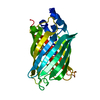
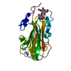
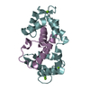
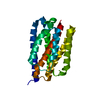
| Title | PSPH Human phosphoserine phosphatase | |||||||||
 Components Components | Phosphoserine phosphatase | |||||||||
 Keywords Keywords | HYDROLASE / phosphoserine phosphatase / homo sapiens | |||||||||
| Function / homology |  Function and homology information Function and homology informationSerine metabolism / phosphoserine phosphatase / L-phosphoserine phosphatase activity / L-serine metabolic process / L-serine biosynthetic process / response to testosterone / response to mechanical stimulus / response to nutrient levels / in utero embryonic development / magnesium ion binding ...Serine metabolism / phosphoserine phosphatase / L-phosphoserine phosphatase activity / L-serine metabolic process / L-serine biosynthetic process / response to testosterone / response to mechanical stimulus / response to nutrient levels / in utero embryonic development / magnesium ion binding / protein homodimerization activity / identical protein binding / cytoplasm / cytosol Similarity search - Function | |||||||||
| Biological species |  Homo sapiens (human) Homo sapiens (human) | |||||||||
| Method |  X-RAY DIFFRACTION / SYNCHROTRON / MOLECULAR REPLACEMENT / Resolution: 1.929 Å X-RAY DIFFRACTION / SYNCHROTRON / MOLECULAR REPLACEMENT / Resolution: 1.929 Å | |||||||||
 Authors Authors | Wouters, J. / Haufroid, M. / Mirgaux, M. | |||||||||
 Citation Citation | Journal: Acta Crystallogr D Struct Biol / Year: 2019 Title: Crystal structures and snapshots along the reaction pathway of human phosphoserine phosphatase. Authors: Haufroid, M. / Mirgaux, M. / Leherte, L. / Wouters, J. | |||||||||
| History |
|
- Structure visualization
| Structure viewer | Molecule: MolmilJmol/JSmol |
|---|
- Downloads & links
-Download
| PDBx/mmCIF format | 6hyj.cif.gz | 105.4 KB | Display | PDBx/mmCIF format |
|---|---|---|---|---|
| PDB format | pdb6hyj.ent.gz | 80.2 KB | Display | PDB format |
| PDBx/mmJSON format | 6hyj.json.gz | Tree view | PDBx/mmJSON format | |
| Others |  Other downloads Other downloads |
-Validation report
| Arichive directory | https://data.pdbj.org/pub/pdb/validation_reports/hy/6hyjftp://data.pdbj.org/pub/pdb/validation_reports/hy/6hyj | HTTPS FTP |
|---|
-Related structure data
| Related structure data | 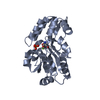 6hyyC  6q6jC  1nnlS S: Starting model for refinement C: citing same article ( |
|---|---|
| Similar structure data |


-Links
 PDBj
PDBj

- Assembly
| Deposited unit | 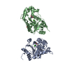
| ||||||||
|---|---|---|---|---|---|---|---|---|---|
| 1 | 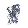
| ||||||||
| 2 | 
| ||||||||
| Unit cell |
|
-Components
| #1: Protein | Mass: 24923.584 Da / Num. of mol.: 2 Source method: isolated from a genetically manipulated source Source: (gene. exp.) Homo sapiens (human) / Gene: PSPH / Production host:  #2: Chemical |   Type: L-peptide linking / Mass: 185.072 Da / Num. of mol.: 2 / Source method: obtained synthetically / Formula: C3H8NO6P Type: L-peptide linking / Mass: 185.072 Da / Num. of mol.: 2 / Source method: obtained synthetically / Formula: C3H8NO6P#3: Chemical |   Mass: 40.078 Da / Num. of mol.: 2 / Source method: obtained synthetically / Formula: Ca Mass: 40.078 Da / Num. of mol.: 2 / Source method: obtained synthetically / Formula: Ca#4: Chemical | ChemComp-SER / |   Type: L-peptide linking / Mass: 105.093 Da / Num. of mol.: 1 / Source method: obtained synthetically / Formula: C3H7NO3 Type: L-peptide linking / Mass: 105.093 Da / Num. of mol.: 1 / Source method: obtained synthetically / Formula: C3H7NO3#5: Water | ChemComp-HOH / |  Mass: 18.015 Da / Num. of mol.: 185 / Source method: isolated from a natural source / Formula: H2O Mass: 18.015 Da / Num. of mol.: 185 / Source method: isolated from a natural source / Formula: H2O |
|---|
-Experimental details
-Experiment
| Experiment | Method: X-RAY DIFFRACTION / Number of used crystals: 1 |
|---|
- Sample preparation
| Crystal | Density Matthews: 2.46 Å3/Da / Density % sol: 50.09 % |
|---|---|
| Crystal grow | Temperature: 293 K / Method: vapor diffusion, hanging drop / pH: 6.5 Details: CaCl2 0.15 M; sodium cacodylate 0.1 M pH 6.5; PEG 2000 20% |
-Data collection
| Diffraction | Mean temperature: 100 K / Serial crystal experiment: N |
|---|---|
| Diffraction source | Source: SYNCHROTRON / Site: SOLEIL  / Beamline: PROXIMA 1 / Wavelength: 0.97857 Å / Beamline: PROXIMA 1 / Wavelength: 0.97857 Å |
| Detector | Type: ADSC QUANTUM 315r / Detector: CCD / Date: Apr 5, 2018 |
| Radiation | Protocol: SINGLE WAVELENGTH / Monochromatic (M) / Laue (L): M / Scattering type: x-ray |
| Radiation wavelength | Wavelength: 0.97857 Å / Relative weight: 1 |
| Reflection | Resolution: 1.929→34.23 Å / Num. obs: 36957 / % possible obs: 98.66 % / Redundancy: 2 % / Biso Wilson estimate: 43.32 Å2 / CC1/2: 0.999 / Rmerge(I) obs: 0.02987 / Rpim(I) all: 0.02987 / Rrim(I) all: 0.04224 / Net I/σ(I): 11.27 |
| Reflection shell | Resolution: 1.929→1.998 Å / Redundancy: 1.9 % / Rmerge(I) obs: 0.5529 / Mean I/σ(I) obs: 0.96 / Num. unique obs: 3228 / CC1/2: 0.589 / Rpim(I) all: 0.5529 / Rrim(I) all: 0.7819 / % possible all: 87.57 |
- Processing
| Software |
| ||||||||||||||||||||||||||||||||||||||||||||||||||||||||||||||||||||||||||||||||||||||||||||||||||
|---|---|---|---|---|---|---|---|---|---|---|---|---|---|---|---|---|---|---|---|---|---|---|---|---|---|---|---|---|---|---|---|---|---|---|---|---|---|---|---|---|---|---|---|---|---|---|---|---|---|---|---|---|---|---|---|---|---|---|---|---|---|---|---|---|---|---|---|---|---|---|---|---|---|---|---|---|---|---|---|---|---|---|---|---|---|---|---|---|---|---|---|---|---|---|---|---|---|---|---|
| Refinement | Method to determine structure: MOLECULAR REPLACEMENT Starting model: 1NNL Resolution: 1.929→34.23 Å / SU ML: 0.35 / Cross valid method: FREE R-VALUE / σ(F): 1.33 / Phase error: 27.8
| ||||||||||||||||||||||||||||||||||||||||||||||||||||||||||||||||||||||||||||||||||||||||||||||||||
| Solvent computation | Shrinkage radii: 0.9 Å / VDW probe radii: 1.11 Å | ||||||||||||||||||||||||||||||||||||||||||||||||||||||||||||||||||||||||||||||||||||||||||||||||||
| Displacement parameters | Biso max: 111.65 Å2 / Biso mean: 48.8391 Å2 / Biso min: 23.74 Å2 | ||||||||||||||||||||||||||||||||||||||||||||||||||||||||||||||||||||||||||||||||||||||||||||||||||
| Refinement step | Cycle: final / Resolution: 1.929→34.23 Å
| ||||||||||||||||||||||||||||||||||||||||||||||||||||||||||||||||||||||||||||||||||||||||||||||||||
| LS refinement shell | Refine-ID: X-RAY DIFFRACTION / Rfactor Rfree error: 0 / Total num. of bins used: 13
|
