 Movie
Movie Controller

[English] 日本語
 Yorodumi
- PDB-6hxe: Crystal structure of psychrophilic phosphoglycerate kinase from P... -
+ Open data

- Basic information
| Entry | Database: PDB / ID: 6hxe | ||||||
|---|---|---|---|---|---|---|---|
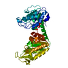
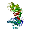
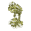
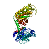
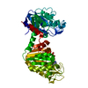
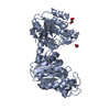
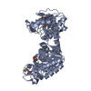
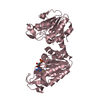
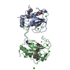
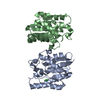
| Title | Crystal structure of psychrophilic phosphoglycerate kinase from Pseudomonas TACII18 in complex with 3-phosphoglycerate | ||||||
 Components Components | Phosphoglycerate kinase | ||||||
 Keywords Keywords | TRANSFERASE / Complex / 3-phosphoglycerate / hinge binding | ||||||
| Function / homology |  Function and homology information Function and homology informationphosphoglycerate kinase / phosphoglycerate kinase activity / glycolytic process / gluconeogenesis / ADP binding / ATP binding / cytosol Similarity search - Function | ||||||
| Biological species |  Pseudomonas sp. 'TAC II 18' (bacteria) Pseudomonas sp. 'TAC II 18' (bacteria) | ||||||
| Method |  X-RAY DIFFRACTION / MOLECULAR REPLACEMENT / Resolution: 2.1 Å X-RAY DIFFRACTION / MOLECULAR REPLACEMENT / Resolution: 2.1 Å | ||||||
 Authors Authors | Mandelman, D. / Haser, R. / Aghajari, N. | ||||||
| Funding support |  France, 1items France, 1items
| ||||||
 Citation Citation | Journal: Extremophiles / Year: 2019 Title: Structural determinants increasing flexibility confer cold adaptation in psychrophilic phosphoglycerate kinase. Authors: Mandelman, D. / Ballut, L. / Wolff, D.A. / Feller, G. / Gerday, C. / Haser, R. / Aghajari, N. #1: Journal: Acta Crystallogr. D Biol. Crystallogr. / Year: 2001 Title: Crystallization and preliminary X-ray analysis of a bacterial psychrophilic enzyme, phosphoglycerate kinase. Authors: Mandelman, D. / Bentahir, M. / Feller, G. / Gerday, C. / Haser, R. | ||||||
| History |
|
- Structure visualization
| Structure viewer | Molecule: MolmilJmol/JSmol |
|---|
- Downloads & links
-Download
| PDBx/mmCIF format | 6hxe.cif.gz | 163.6 KB | Display | PDBx/mmCIF format |
|---|---|---|---|---|
| PDB format | pdb6hxe.ent.gz | 127.9 KB | Display | PDB format |
| PDBx/mmJSON format | 6hxe.json.gz | Tree view | PDBx/mmJSON format | |
| Others |  Other downloads Other downloads |
-Validation report
| Arichive directory | https://data.pdbj.org/pub/pdb/validation_reports/hx/6hxeftp://data.pdbj.org/pub/pdb/validation_reports/hx/6hxe | HTTPS FTP |
|---|
-Related structure data
| Related structure data |  6i06C 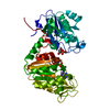 1vpeS C: citing same article ( S: Starting model for refinement |
|---|---|
| Similar structure data |
-Links
 PDBj
PDBj
- Assembly
| Deposited unit | 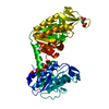
| ||||||||
|---|---|---|---|---|---|---|---|---|---|
| 1 |
| ||||||||
| Unit cell |
|
-Components
| #1: Protein | Mass: 40267.207 Da / Num. of mol.: 1 Source method: isolated from a genetically manipulated source Details: There is a mismatch between the sequence deposited in uniprot (ID: Q9RBS3) and the structure at position 150. This should clearly be a Ser as judged from the electron density and not a Pro ...Details: There is a mismatch between the sequence deposited in uniprot (ID: Q9RBS3) and the structure at position 150. This should clearly be a Ser as judged from the electron density and not a Pro as listed in the sequence. Idem for residue 219 which is not a Ser but an Asp and residue 358 which is not a Tyr but a Gln. Following residues have been refined as alanines due to missing electron density for the side-chains: 27, 102, 243, 248, 268, 292, 327, 348 Source: (gene. exp.) Pseudomonas sp. 'TAC II 18' (bacteria) / Gene: pgk / Production host: |
|---|---|
| #2: Chemical | ChemComp-3PG /   Mass: 186.057 Da / Num. of mol.: 1 / Source method: obtained synthetically / Formula: C3H7O7P Mass: 186.057 Da / Num. of mol.: 1 / Source method: obtained synthetically / Formula: C3H7O7P |
| #3: Water | ChemComp-HOH /  Mass: 18.015 Da / Num. of mol.: 267 / Source method: isolated from a natural source / Formula: H2O Mass: 18.015 Da / Num. of mol.: 267 / Source method: isolated from a natural source / Formula: H2O |
-Experimental details
-Experiment
| Experiment | Method: X-RAY DIFFRACTION / Number of used crystals: 1 |
|---|
- Sample preparation
| Crystal | Density Matthews: 2.09 Å3/Da / Density % sol: 41.24 % |
|---|---|
| Crystal grow | Temperature: 290 K / Method: vapor diffusion, hanging drop / pH: 8.4 Details: 30% polyethylene glycol (PEG) 4000, 0.2 M MgCl2 and 0.1 M Tris |
-Data collection
| Diffraction | Mean temperature: 100 K / Serial crystal experiment: N |
|---|---|
| Diffraction source | Source: ROTATING ANODE / Type: RIGAKU / Wavelength: 1.5418 Å |
| Detector | Type: MARRESEARCH / Detector: IMAGE PLATE / Date: Nov 10, 1999 |
| Radiation | Monochromator: Osmic mirrors / Protocol: SINGLE WAVELENGTH / Monochromatic (M) / Laue (L): M / Scattering type: x-ray |
| Radiation wavelength | Wavelength: 1.5418 Å / Relative weight: 1 |
| Reflection | Resolution: 2.1→46 Å / Num. obs: 61709 / % possible obs: 99.4 % / Redundancy: 3.3 % / Net I/σ(I): 8.6 |
| Reflection shell | Resolution: 2.1→2.18 Å |
- Processing
| Software |
| ||||||||||||||||||||||||||||||||||||||||||||||||||||||||||||||||||||||
|---|---|---|---|---|---|---|---|---|---|---|---|---|---|---|---|---|---|---|---|---|---|---|---|---|---|---|---|---|---|---|---|---|---|---|---|---|---|---|---|---|---|---|---|---|---|---|---|---|---|---|---|---|---|---|---|---|---|---|---|---|---|---|---|---|---|---|---|---|---|---|---|
| Refinement | Method to determine structure: MOLECULAR REPLACEMENT Starting model: 1vpe Resolution: 2.1→16.887 Å / SU ML: 0.26 / Cross valid method: FREE R-VALUE / σ(F): 1.97 / Phase error: 25.52
| ||||||||||||||||||||||||||||||||||||||||||||||||||||||||||||||||||||||
| Solvent computation | Shrinkage radii: 0.9 Å / VDW probe radii: 1.11 Å | ||||||||||||||||||||||||||||||||||||||||||||||||||||||||||||||||||||||
| Refinement step | Cycle: LAST / Resolution: 2.1→16.887 Å
| ||||||||||||||||||||||||||||||||||||||||||||||||||||||||||||||||||||||
| Refine LS restraints |
| ||||||||||||||||||||||||||||||||||||||||||||||||||||||||||||||||||||||
| LS refinement shell |
| ||||||||||||||||||||||||||||||||||||||||||||||||||||||||||||||||||||||
| Refinement TLS params. | Method: refined / Origin x: 55.225 Å / Origin y: 6.5017 Å / Origin z: 0.8257 Å
| ||||||||||||||||||||||||||||||||||||||||||||||||||||||||||||||||||||||
| Refinement TLS group | Selection details: all |
