 Movie
Movie Controller

[English] 日本語
 Yorodumi
- PDB-6hwl: Glucosamine kinase in complex with glucosamine, ADP and inorganic... -
+ Open data

- Basic information
| Entry | Database: PDB / ID: 6hwl | ||||||
|---|---|---|---|---|---|---|---|
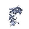
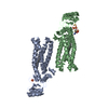
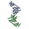
| Title | Glucosamine kinase in complex with glucosamine, ADP and inorganic phosphate | ||||||
 Components Components | Glucosamine kinase | ||||||
 Keywords Keywords | TRANSFERASE / Glucosamine kinase | ||||||
| Function / homology |  Function and homology information Function and homology informationglucosamine kinase / glucosamine kinase activity / carbohydrate metabolic process / magnesium ion binding / ATP binding Similarity search - Function | ||||||
| Biological species |  Streptacidiphilus jiangxiensis (bacteria) Streptacidiphilus jiangxiensis (bacteria) | ||||||
| Method |  X-RAY DIFFRACTION / SYNCHROTRON / MOLECULAR REPLACEMENT / Resolution: 2.148 Å X-RAY DIFFRACTION / SYNCHROTRON / MOLECULAR REPLACEMENT / Resolution: 2.148 Å | ||||||
 Authors Authors | Manso, J.A. / Pereira, P.J.B. | ||||||
 Citation Citation | Journal: mBio / Year: 2019 Title: Molecular Fingerprints for a Novel Enzyme Family in with Glucosamine Kinase Activity. Authors: José A Manso / Daniela Nunes-Costa / Sandra Macedo-Ribeiro / Nuno Empadinhas / Pedro José Barbosa Pereira /  Abstract: have long been the main source of antibiotics, secondary metabolites with tightly controlled biosynthesis by environmental and physiological factors. Phosphorylation of exogenous glucosamine has ... have long been the main source of antibiotics, secondary metabolites with tightly controlled biosynthesis by environmental and physiological factors. Phosphorylation of exogenous glucosamine has been suggested as a mechanism for incorporation of this extracellular material into secondary metabolite biosynthesis, but experimental evidence of specific glucosamine kinases in is lacking. Here, we present the molecular fingerprints for the identification of a unique family of actinobacterial glucosamine kinases. Structural and biochemical studies on a distinctive kinase from the soil bacterium unveiled its preference for glucosamine and provided structural evidence of a phosphoryl transfer to this substrate. Conservation of glucosamine-contacting residues across a large number of uncharacterized actinobacterial proteins unveiled a specific glucosamine binding sequence motif. This family of kinases and their genetic context may represent the missing link for the incorporation of environmental glucosamine into the antibiotic biosynthesis pathways in and can be explored to enhance antibiotic production. The discovery of novel enzymes involved in antibiotic biosynthesis pathways is currently a topic of utmost importance. The high levels of antibiotic resistance detected worldwide threaten our ability to combat infections and other 20th-century medical achievements, namely, organ transplantation or cancer chemotherapy. We have identified and characterized a unique family of enzymes capable of phosphorylating glucosamine to glucosamine-6-phosphate, a crucial molecule directly involved in the activation of antibiotic production pathways in , nature's main source of antimicrobials. The consensus sequence identified for these glucosamine kinases will help establish a molecular fingerprint to reveal yet-uncharacterized sequences in antibiotic producers, which should have an important impact in biotechnological and biomedical applications, including the enhancement and optimization of antibiotic production. | ||||||
| History |
|
- Structure visualization
| Structure viewer | Molecule: MolmilJmol/JSmol |
|---|
- Downloads & links
-Download
| PDBx/mmCIF format | 6hwl.cif.gz | 321.4 KB | Display | PDBx/mmCIF format |
|---|---|---|---|---|
| PDB format | pdb6hwl.ent.gz | 260.3 KB | Display | PDB format |
| PDBx/mmJSON format | 6hwl.json.gz | Tree view | PDBx/mmJSON format | |
| Others |  Other downloads Other downloads |
-Validation report
| Arichive directory | https://data.pdbj.org/pub/pdb/validation_reports/hw/6hwlftp://data.pdbj.org/pub/pdb/validation_reports/hw/6hwl | HTTPS FTP |
|---|
-Related structure data
| Related structure data |  6hwjC 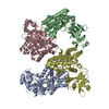 6hwkC C: citing same article ( |
|---|---|
| Similar structure data | |
| Experimental dataset #1 | Data reference: 10.15785/SBGRID/616 / Data set type: diffraction image data |


-Links
 PDBj
PDBj- Assembly
| Deposited unit | 
| ||||||||
|---|---|---|---|---|---|---|---|---|---|
| 1 |
| ||||||||
| Unit cell |
|
-Components
-Protein / Sugars , 2 types, 3 molecules AB
| #1: Protein | Mass: 48308.703 Da / Num. of mol.: 2 Source method: isolated from a genetically manipulated source Source: (gene. exp.) Streptacidiphilus jiangxiensis (bacteria)Gene: SAMN05414137_114149 / Production host: #8: Sugar | ChemComp-GCS / |  Type: D-saccharide, beta linking / Mass: 179.171 Da / Num. of mol.: 1 Type: D-saccharide, beta linking / Mass: 179.171 Da / Num. of mol.: 1Source method: isolated from a genetically manipulated source Formula: C6H13NO5 |
|---|
-Non-polymers , 7 types, 253 molecules 


| #2: Chemical |  Mass: 35.453 Da / Num. of mol.: 2 / Source method: obtained synthetically / Formula: Cl Mass: 35.453 Da / Num. of mol.: 2 / Source method: obtained synthetically / Formula: Cl#3: Chemical | ChemComp-BTB / |  Mass: 209.240 Da / Num. of mol.: 1 / Source method: obtained synthetically / Formula: C8H19NO5 / Comment: pH buffer*YM Mass: 209.240 Da / Num. of mol.: 1 / Source method: obtained synthetically / Formula: C8H19NO5 / Comment: pH buffer*YM#4: Chemical |  Mass: 106.120 Da / Num. of mol.: 3 / Source method: obtained synthetically / Formula: C4H10O3 Mass: 106.120 Da / Num. of mol.: 3 / Source method: obtained synthetically / Formula: C4H10O3#5: Chemical | ChemComp-ADP / |  Mass: 427.201 Da / Num. of mol.: 1 / Source method: obtained synthetically / Formula: C10H15N5O10P2 / Comment: ADP, energy-carrying molecule*YM Mass: 427.201 Da / Num. of mol.: 1 / Source method: obtained synthetically / Formula: C10H15N5O10P2 / Comment: ADP, energy-carrying molecule*YM#6: Chemical |  Mass: 24.305 Da / Num. of mol.: 2 / Source method: obtained synthetically / Formula: Mg Mass: 24.305 Da / Num. of mol.: 2 / Source method: obtained synthetically / Formula: Mg#7: Chemical | ChemComp-PO4 / |  Mass: 94.971 Da / Num. of mol.: 1 / Source method: obtained synthetically / Formula: PO4 Mass: 94.971 Da / Num. of mol.: 1 / Source method: obtained synthetically / Formula: PO4#9: Water | ChemComp-HOH / | Mass: 18.015 Da / Num. of mol.: 243 / Source method: isolated from a natural source / Formula: H2O |
|---|
-Experimental details
-Experiment
| Experiment | Method: X-RAY DIFFRACTION / Number of used crystals: 1 |
|---|
- Sample preparation
| Crystal | Density Matthews: 2.27 Å3/Da / Density % sol: 45.84 % |
|---|---|
| Crystal grow | Temperature: 293 K / Method: vapor diffusion, sitting drop Details: 100 mM Bis-Tris pH 6.1, 16% (wt/vol) PEG 3350, 0.15 M MgCl2 |
-Data collection
| Diffraction | Mean temperature: 100 K / Serial crystal experiment: N |
|---|---|
| Diffraction source | Source: SYNCHROTRON / Site: ESRF  / Beamline: MASSIF-3 / Wavelength: 0.9677 Å / Beamline: MASSIF-3 / Wavelength: 0.9677 Å |
| Detector | Type: DECTRIS EIGER X 4M / Detector: PIXEL / Date: Mar 2, 2017 |
| Radiation | Protocol: SINGLE WAVELENGTH / Monochromatic (M) / Laue (L): M / Scattering type: x-ray |
| Radiation wavelength | Wavelength: 0.9677 Å / Relative weight: 1 |
| Reflection | Resolution: 2.148→60.175 Å / Num. obs: 46229 / % possible obs: 98.3 % / Redundancy: 7 % / Biso Wilson estimate: 40.8 Å2 / CC1/2: 0.998 / Rmerge(I) obs: 0.113 / Rpim(I) all: 0.045 / Rrim(I) all: 0.122 / Net I/σ(I): 11.8 |
| Reflection shell | Resolution: 2.15→2.19 Å / Redundancy: 7.3 % / Rmerge(I) obs: 1.543 / Mean I/σ(I) obs: 1.3 / Num. unique obs: 2340 / CC1/2: 0.559 / Rpim(I) all: 0.608 / Rrim(I) all: 1.66 / % possible all: 99.2 |
- Processing
| Software |
| ||||||||||||||||||||||||||||||||||||||||||||||||||||||||||||||||||||||||||||||||||||||||||||||||||||||||||||||||||||||||||||||
|---|---|---|---|---|---|---|---|---|---|---|---|---|---|---|---|---|---|---|---|---|---|---|---|---|---|---|---|---|---|---|---|---|---|---|---|---|---|---|---|---|---|---|---|---|---|---|---|---|---|---|---|---|---|---|---|---|---|---|---|---|---|---|---|---|---|---|---|---|---|---|---|---|---|---|---|---|---|---|---|---|---|---|---|---|---|---|---|---|---|---|---|---|---|---|---|---|---|---|---|---|---|---|---|---|---|---|---|---|---|---|---|---|---|---|---|---|---|---|---|---|---|---|---|---|---|---|---|
| Refinement | Method to determine structure: MOLECULAR REPLACEMENT Starting model: D_1200012387 Resolution: 2.148→39.888 Å / SU ML: 0.27 / Cross valid method: FREE R-VALUE / σ(F): 1.36 / Phase error: 25.71
| ||||||||||||||||||||||||||||||||||||||||||||||||||||||||||||||||||||||||||||||||||||||||||||||||||||||||||||||||||||||||||||||
| Solvent computation | Shrinkage radii: 0.9 Å / VDW probe radii: 1.11 Å | ||||||||||||||||||||||||||||||||||||||||||||||||||||||||||||||||||||||||||||||||||||||||||||||||||||||||||||||||||||||||||||||
| Refinement step | Cycle: LAST / Resolution: 2.148→39.888 Å
| ||||||||||||||||||||||||||||||||||||||||||||||||||||||||||||||||||||||||||||||||||||||||||||||||||||||||||||||||||||||||||||||
| Refine LS restraints |
| ||||||||||||||||||||||||||||||||||||||||||||||||||||||||||||||||||||||||||||||||||||||||||||||||||||||||||||||||||||||||||||||
| LS refinement shell |
| ||||||||||||||||||||||||||||||||||||||||||||||||||||||||||||||||||||||||||||||||||||||||||||||||||||||||||||||||||||||||||||||
| Refinement TLS params. | Method: refined / Refine-ID: X-RAY DIFFRACTION
| ||||||||||||||||||||||||||||||||||||||||||||||||||||||||||||||||||||||||||||||||||||||||||||||||||||||||||||||||||||||||||||||
| Refinement TLS group |
|
