 Movie
Movie Controller

[English] 日本語
 Yorodumi
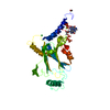
- PDB-6hw2: The Crystal Structure of CaV beta4c in complex with HP1gamma chro... -
+ Open data

- Basic information
| Entry | Database: PDB / ID: 6hw2 | |||||||||
|---|---|---|---|---|---|---|---|---|---|---|
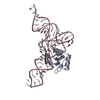
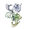
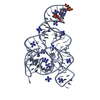
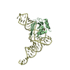
| Title | The Crystal Structure of CaV beta4c in complex with HP1gamma chromo shadow domains | |||||||||
 Components Components |
| |||||||||
 Keywords Keywords | TRANSPORT PROTEIN / Complex | |||||||||
| Function / homology |  Function and homology information Function and homology informationsenescence-associated heterochromatin focus / voltage-gated calcium channel activity involved in regulation of presynaptic cytosolic calcium levels / chromatin lock complex / positive regulation of protein localization to nucleolus / Presynaptic depolarization and calcium channel opening / gamma-aminobutyric acid secretion / high voltage-gated calcium channel activity / detection of light stimulus involved in visual perception / cAMP metabolic process / histone methyltransferase binding ...senescence-associated heterochromatin focus / voltage-gated calcium channel activity involved in regulation of presynaptic cytosolic calcium levels / chromatin lock complex / positive regulation of protein localization to nucleolus / Presynaptic depolarization and calcium channel opening / gamma-aminobutyric acid secretion / high voltage-gated calcium channel activity / detection of light stimulus involved in visual perception / cAMP metabolic process / histone methyltransferase binding / Peyer's patch development / condensed chromosome, centromeric region / muscle cell development / neuronal action potential propagation / nervous system process / nuclear inner membrane / adult walking behavior / voltage-gated calcium channel complex / : / negative regulation of G1/S transition of mitotic cell cycle / gamma-aminobutyric acid signaling pathway / neuromuscular junction development / regulation of synaptic vesicle exocytosis / Transcriptional Regulation by E2F6 / chromosome, centromeric region / cellular response to dexamethasone stimulus / site of DNA damage / pericentric heterochromatin / voltage-gated calcium channel activity / spleen development / heterochromatin / thymus development / cellular response to leukemia inhibitory factor / transcription coregulator binding / ERCC6 (CSB) and EHMT2 (G9a) positively regulate rRNA expression / synaptic transmission, glutamatergic / regulation of membrane potential / RNA Polymerase I Promoter Escape / euchromatin / RNA polymerase II transcription regulator complex / spindle / calcium ion transport / rhythmic process / nuclear envelope / heterochromatin formation / T cell receptor signaling pathway / presynapse / chemical synaptic transmission / chromosome, telomeric region / nuclear speck / chromatin remodeling / protein domain specific binding / negative regulation of cell population proliferation / negative regulation of DNA-templated transcription / DNA damage response / chromatin binding / protein kinase binding / chromatin / glutamatergic synapse / enzyme binding / nucleoplasm / identical protein binding / nucleus / plasma membrane Similarity search - Function | |||||||||
| Biological species |   Homo sapiens (human) Homo sapiens (human) | |||||||||
| Method |  X-RAY DIFFRACTION / SYNCHROTRON / MOLECULAR REPLACEMENT / Resolution: 1.941 Å X-RAY DIFFRACTION / SYNCHROTRON / MOLECULAR REPLACEMENT / Resolution: 1.941 Å | |||||||||
 Authors Authors | Tanner, N. / Tripathy, D.R. / Hirsch, J.A. | |||||||||
| Funding support |  Israel, 2items Israel, 2items
| |||||||||
 Citation Citation | Journal: To Be Published Title: The Crystal Structure of CaV beta4c in complex with HP1gamma chromo shadow domains Authors: Tanner, N. / Tripathy, D.R. / Hirsch, J.A. | |||||||||
| History |
|
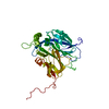
- Structure visualization
| Structure viewer | Molecule: MolmilJmol/JSmol |
|---|
- Downloads & links
-Download
| PDBx/mmCIF format | 6hw2.cif.gz | 175.8 KB | Display | PDBx/mmCIF format |
|---|---|---|---|---|
| PDB format | pdb6hw2.ent.gz | 135.1 KB | Display | PDB format |
| PDBx/mmJSON format | 6hw2.json.gz | Tree view | PDBx/mmJSON format | |
| Others |  Other downloads Other downloads |
-Validation report
| Arichive directory | https://data.pdbj.org/pub/pdb/validation_reports/hw/6hw2ftp://data.pdbj.org/pub/pdb/validation_reports/hw/6hw2 | HTTPS FTP |
|---|
-Related structure data
| Related structure data |  1vyvS S: Starting model for refinement |
|---|---|
| Similar structure data |


-Links
 PDBj
PDBj


- Assembly
| Deposited unit | 
| ||||||||||||
|---|---|---|---|---|---|---|---|---|---|---|---|---|---|
| 1 |
| ||||||||||||
| Unit cell |
|
-Components
-Protein , 2 types, 3 molecules ABC
| #1: Protein | Mass: 17531.834 Da / Num. of mol.: 1 Source method: isolated from a genetically manipulated source Source: (gene. exp.) Production host:  References: UniProt: D4A055*PLUS |
|---|---|
| #2: Protein | Mass: 8468.519 Da / Num. of mol.: 2 Source method: isolated from a genetically manipulated source Source: (gene. exp.) Homo sapiens (human) / Gene: CBX3Production host: References: UniProt: Q13185 |
-Non-polymers , 5 types, 213 molecules 


| #3: Chemical | ChemComp-CA /  Mass: 40.078 Da / Num. of mol.: 4 / Source method: obtained synthetically / Formula: Ca Mass: 40.078 Da / Num. of mol.: 4 / Source method: obtained synthetically / Formula: Ca#4: Chemical | ChemComp-GOL / |  Mass: 92.094 Da / Num. of mol.: 1 / Source method: obtained synthetically / Formula: C3H8O3 Mass: 92.094 Da / Num. of mol.: 1 / Source method: obtained synthetically / Formula: C3H8O3#5: Chemical | ChemComp-BME /  Mass: 78.133 Da / Num. of mol.: 5 / Source method: obtained synthetically / Formula: C2H6OS Mass: 78.133 Da / Num. of mol.: 5 / Source method: obtained synthetically / Formula: C2H6OS#6: Chemical | ChemComp-ACT / |  Mass: 59.044 Da / Num. of mol.: 1 / Source method: obtained synthetically / Formula: C2H3O2 Mass: 59.044 Da / Num. of mol.: 1 / Source method: obtained synthetically / Formula: C2H3O2#7: Water | ChemComp-HOH / | Mass: 18.015 Da / Num. of mol.: 202 / Source method: isolated from a natural source / Formula: H2O |
|---|
-Details
| Has protein modification | Y |
|---|
-Experimental details
-Experiment
| Experiment | Method: X-RAY DIFFRACTION / Number of used crystals: 1 |
|---|
- Sample preparation
| Crystal | Density Matthews: 2.63 Å3/Da / Density % sol: 53.17 % |
|---|---|
| Crystal grow | Temperature: 292 K / Method: vapor diffusion, hanging drop / pH: 5 Details: 0.15 M NaCl, 12-14% PEG 3350 , pH 5.0, calcium acetate, VAPOR DIFFUSION, temperature 292K |
-Data collection
| Diffraction | Mean temperature: 100 K / Serial crystal experiment: N |
|---|---|
| Diffraction source | Source: SYNCHROTRON / Site: ESRF  / Beamline: ID14-2 / Wavelength: 0.933 Å / Beamline: ID14-2 / Wavelength: 0.933 Å |
| Detector | Type: ADSC QUANTUM 4 / Detector: CCD / Date: May 17, 2007 |
| Radiation | Protocol: SINGLE WAVELENGTH / Monochromatic (M) / Laue (L): M / Scattering type: x-ray |
| Radiation wavelength | Wavelength: 0.933 Å / Relative weight: 1 |
| Reflection | Resolution: 1.941→33.12 Å / Num. obs: 26484 / % possible obs: 99.8 % / Redundancy: 5.2 % / Rmerge(I) obs: 0.047 / Net I/σ(I): 12.8 |
| Reflection shell | Resolution: 1.941→2.01 Å / Rmerge(I) obs: 0.404 / Num. unique obs: 2518 |
- Processing
| Software |
| ||||||||||||||||
|---|---|---|---|---|---|---|---|---|---|---|---|---|---|---|---|---|---|
| Refinement | Method to determine structure: MOLECULAR REPLACEMENT Starting model: 1VYV Resolution: 1.941→33.12 Å / Cross valid method: FREE R-VALUE
| ||||||||||||||||
| Refinement step | Cycle: LAST / Resolution: 1.941→33.12 Å
| ||||||||||||||||
| LS refinement shell | Resolution: 1.941→2.0104 Å
|
