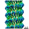
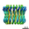
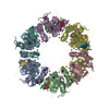
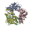
Journal: Proc Natl Acad Sci U S A / Year: 2019 Title: Ambidextrous helical nanotubes from self-assembly of designed helical hairpin motifs. Authors: Spencer A Hughes / Fengbin Wang / Shengyuan Wang / Mark A B Kreutzberger / Tomasz Osinski / Albina Orlova / Joseph S Wall / Xiaobing Zuo / Edward H Egelman / Vincent P Conticello / Abstract: Tandem repeat proteins exhibit native designability and represent potentially useful scaffolds for the construction of synthetic biomimetic assemblies. We have designed 2 synthetic peptides, HEAT_R1 ...Tandem repeat proteins exhibit native designability and represent potentially useful scaffolds for the construction of synthetic biomimetic assemblies. We have designed 2 synthetic peptides, HEAT_R1 and LRV_M3Δ1, based on the consensus sequences of single repeats of thermophilic HEAT (PBS_HEAT) and Leucine-Rich Variant (LRV) structural motifs, respectively. Self-assembly of the peptides afforded high-aspect ratio helical nanotubes. Cryo-electron microscopy with direct electron detection was employed to analyze the structures of the solvated filaments. The 3D reconstructions from the cryo-EM maps led to atomic models for the HEAT_R1 and LRV_M3Δ1 filaments at resolutions of 6.0 and 4.4 Å, respectively. Surprisingly, despite sequence similarity at the lateral packing interface, HEAT_R1 and LRV_M3Δ1 filaments adopt the opposite helical hand and differ significantly in helical geometry, while retaining a local conformation similar to previously characterized repeat proteins of the same class. The differences in the 2 filaments could be rationalized on the basis of differences in cohesive interactions at the lateral and axial interfaces. These structural data reinforce previous observations regarding the structural plasticity of helical protein assemblies and the need for high-resolution structural analysis. Despite these observations, the native designability of tandem repeat proteins offers the opportunity to engineer novel helical nanotubes. Moreover, the resultant nanotubes have independently addressable and chemically distinguishable interior and exterior surfaces that would facilitate applications in selective recognition, transport, and release.
Resolution: 4.4 Å / Resolution method: OTHER / Num. of particles: 62616 / Algorithm: BACK PROJECTION / Details: Model:Map FSC 0.38 cut off, d99 and d model / Symmetry type: HELICAL
+
About Yorodumi
-
News
-
Feb 9, 2022. New format data for meta-information of EMDB entries
New format data for meta-information of EMDB entries
Version 3 of the EMDB header file is now the official format.
The previous official version 1.9 will be removed from the archive.
In the structure databanks used in Yorodumi, some data are registered as the other names, "COVID-19 virus" and "2019-nCoV". Here are the details of the virus and the list of structure data.
Jan 31, 2019. EMDB accession codes are about to change! (news from PDBe EMDB page)
EMDB accession codes are about to change! (news from PDBe EMDB page)
The allocation of 4 digits for EMDB accession codes will soon come to an end. Whilst these codes will remain in use, new EMDB accession codes will include an additional digit and will expand incrementally as the available range of codes is exhausted. The current 4-digit format prefixed with “EMD-” (i.e. EMD-XXXX) will advance to a 5-digit format (i.e. EMD-XXXXX), and so on. It is currently estimated that the 4-digit codes will be depleted around Spring 2019, at which point the 5-digit format will come into force.
The EM Navigator/Yorodumi systems omit the EMD- prefix.
Related info.:Q: What is EMD? / ID/Accession-code notation in Yorodumi/EM Navigator
Yorodumi is a browser for structure data from EMDB, PDB, SASBDB, etc.
This page is also the successor to EM Navigator detail page, and also detail information page/front-end page for Omokage search.
The word "yorodu" (or yorozu) is an old Japanese word meaning "ten thousand". "mi" (miru) is to see.
Related info.:EMDB / PDB / SASBDB / Comparison of 3 databanks / Yorodumi Search / Aug 31, 2016. New EM Navigator & Yorodumi / Yorodumi Papers / Jmol/JSmol / Function and homology information / Changes in new EM Navigator and Yorodumi
 Movie
Movie Controller


 Open data
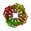
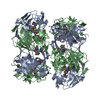
 Basic information
 Components
 Keywords
 Azotobacter vinelandii (bacteria)
 Authors
 United States, 1items
  Citation
 Structure visualization
 Movie viewer
 Downloads & links
 Other downloads


 PDBj
PDBj
 Assembly
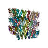
 Sample preparation
 Electron microscopy imaging
 FIELD EMISSION GUN / Accelerating voltage: 300 kV / Illumination mode: FLOOD BEAM
 Processing