 Movie
Movie Controller

[English] 日本語
 Yorodumi
- EMDB-3546: Structure of a novel N-type ATPase rotor ring (c17) from Burkhold... -
+ Open data

- Basic information
| Entry | Database: EMDB / ID: EMD-3546 | |||||||||
|---|---|---|---|---|---|---|---|---|---|---|
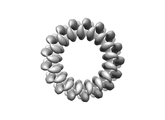
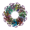
| Title | Structure of a novel N-type ATPase rotor ring (c17) from Burkholderia pseudomallei | |||||||||
 Map data Map data | ||||||||||
 Sample Sample |
| |||||||||
| Biological species |  Burkholderia pseudomallei (bacteria) Burkholderia pseudomallei (bacteria) | |||||||||
| Method | single particle reconstruction / cryo EM / Resolution: 6.1 Å | |||||||||
 Authors Authors | Wilkes M / Schulz S / Mills DJ / Kuhlbrandt W / Meier T | |||||||||
 Citation Citation | Journal: EMBO Rep / Year: 2017 Title: Molecular architecture of the N-type ATPase rotor ring from . Authors: Sarah Schulz / Martin Wilkes / Deryck J Mills / Werner Kühlbrandt / Thomas Meier /  Abstract: The genome of the highly infectious bacterium harbors an operon that encodes an N-type rotary ATPase, in addition to an operon for a regular F-type rotary ATPase. The molecular architecture of N- ...The genome of the highly infectious bacterium harbors an operon that encodes an N-type rotary ATPase, in addition to an operon for a regular F-type rotary ATPase. The molecular architecture of N-type ATPases is unknown and their biochemical properties and cellular functions are largely unexplored. We studied the NN-type ATPase and investigated the structure and ion specificity of its membrane-embedded c-ring rotor by single-particle electron cryo-microscopy. Of several amphiphilic compounds tested for solubilizing the complex, the choice of the low-density, low-CMC detergent LDAO was optimal in terms of map quality and resolution. The cryoEM map of the c-ring at 6.1 Å resolution reveals a heptadecameric oligomer with a molecular mass of ~141 kDa. Biochemical measurements indicate that the c ring is H specific, demonstrating that the ATPase is proton-coupled. The c ring stoichiometry results in a very high ion-to-ATP ratio of 5.7. We propose that this N-ATPase is a highly efficient proton pump that helps these melioidosis-causing bacteria to survive in the hostile, acidic environment of phagosomes. | |||||||||
| History |
|
- Structure visualization
| Movie |
 Movie viewer Movie viewer |
|---|---|
| Structure viewer | EM map: SurfViewMolmilJmol/JSmol |
| Supplemental images |
- Downloads & links
-EMDB archive
| Map data | emd_3546.map.gz | 7.9 MB | EMDB map data format | |
|---|---|---|---|---|
| Header (meta data) | emd-3546-v30.xmlemd-3546.xml | 8.5 KB 8.5 KB | Display Display | EMDB header |
| Images |  emd_3546.png emd_3546.png | 42.9 KB | ||
| Archive directory |  http://ftp.pdbj.org/pub/emdb/structures/EMD-3546ftp://ftp.pdbj.org/pub/emdb/structures/EMD-3546 http://ftp.pdbj.org/pub/emdb/structures/EMD-3546ftp://ftp.pdbj.org/pub/emdb/structures/EMD-3546 | HTTPS FTP |
-Related structure data
| Similar structure data |
|---|

-Links
| EMDB pages | EMDB (EBI/PDBe) / EMDataResource |
|---|
-Map
| File | Download / File: emd_3546.map.gz / Format: CCP4 / Size: 10.5 MB / Type: IMAGE STORED AS FLOATING POINT NUMBER (4 BYTES) | ||||||||||||||||||||||||||||||||||||||||||||||||||||||||||||
|---|---|---|---|---|---|---|---|---|---|---|---|---|---|---|---|---|---|---|---|---|---|---|---|---|---|---|---|---|---|---|---|---|---|---|---|---|---|---|---|---|---|---|---|---|---|---|---|---|---|---|---|---|---|---|---|---|---|---|---|---|---|
| Projections & slices | Image control
Images are generated by Spider. | ||||||||||||||||||||||||||||||||||||||||||||||||||||||||||||
| Voxel size | X=Y=Z: 1.14 Å | ||||||||||||||||||||||||||||||||||||||||||||||||||||||||||||
| Density |
| ||||||||||||||||||||||||||||||||||||||||||||||||||||||||||||
| Symmetry | Space group: 1 | ||||||||||||||||||||||||||||||||||||||||||||||||||||||||||||
| Details | EMDB XML:
CCP4 map header:
| ||||||||||||||||||||||||||||||||||||||||||||||||||||||||||||
 Z (Sec.)
 Y (Row.)
 X (Col.)


-Supplemental data
- Sample components
-Entire : Rotor ring of the N-type ATPase from B. pseudomallei
| Entire | Name: Rotor ring of the N-type ATPase from B. pseudomallei |
|---|---|
| Components |
|
-Supramolecule #1: Rotor ring of the N-type ATPase from B. pseudomallei
| Supramolecule | Name: Rotor ring of the N-type ATPase from B. pseudomallei / type: complex / ID: 1 / Parent: 0 Details: We determined the molecular architecture of the N-type ATPase rotor ring in the melioidosis-causing bacterium Burkholderia pseudomallei by electron cryo-microscopy. The structure shows an ...Details: We determined the molecular architecture of the N-type ATPase rotor ring in the melioidosis-causing bacterium Burkholderia pseudomallei by electron cryo-microscopy. The structure shows an unusually large c-ring rotor with 17 subunits that, in the acidic environment of the host phagosome, is driven predominantly by protons. |
|---|---|
| Source (natural) | Organism: Burkholderia pseudomallei (bacteria) |
| Recombinant expression | Organism: |
| Molecular weight | Experimental: 8.3 kDa/nm |
-Experimental details
-Structure determination
| Method | cryo EM |
|---|---|
 Processing Processing | single particle reconstruction |
| Aggregation state | particle |
-Sample preparation
| Buffer | pH: 7.5 |
|---|---|
| Grid | Model: Quantifoil R2/2 / Material: COPPER |
| Vitrification | Cryogen name: ETHANE / Chamber humidity: 100 % / Chamber temperature: 283 K / Instrument: FEI VITROBOT MARK III |
- Electron microscopy
| Microscope | JEOL 3200FSC |
|---|---|
| Image recording | Film or detector model: GATAN K2 SUMMIT (4k x 4k) / Average electron dose: 1.8 e/Å2 |
| Electron beam | Acceleration voltage: 300 kV / Electron source:  FIELD EMISSION GUN FIELD EMISSION GUN |
| Electron optics | Illumination mode: FLOOD BEAM / Imaging mode: BRIGHT FIELD / Cs: 4.1 mm |
-Image processing
| Final reconstruction | Applied symmetry - Point group: C17 (17 fold cyclic) / Algorithm: BACK PROJECTION / Resolution.type: BY AUTHOR / Resolution: 6.1 Å / Resolution method: FSC 0.143 CUT-OFF / Number images used: 47000 |
|---|---|
| Initial angle assignment | Type: PROJECTION MATCHING |
| Final angle assignment | Type: PROJECTION MATCHING |
