 ムービー
ムービー コントローラー


+ データを開く

- 基本情報
基本情報
| 登録情報 | データベース: EMDB / ID: EMD-3546 | |||||||||
|---|---|---|---|---|---|---|---|---|---|---|
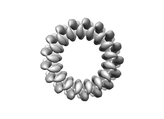
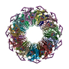
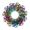
| タイトル | Structure of a novel N-type ATPase rotor ring (c17) from Burkholderia pseudomallei | |||||||||
 マップデータ マップデータ | ||||||||||
 試料 試料 |
| |||||||||
| 生物種 |   Burkholderia pseudomallei (類鼻疽菌) Burkholderia pseudomallei (類鼻疽菌) | |||||||||
| 手法 | 単粒子再構成法 / クライオ電子顕微鏡法 / 解像度: 6.1 Å | |||||||||
 データ登録者 データ登録者 | Wilkes M / Schulz S / Mills DJ / Kuhlbrandt W / Meier T | |||||||||
 引用 引用 | ジャーナル: EMBO Rep / 年: 2017 タイトル: Molecular architecture of the N-type ATPase rotor ring from . 著者: Sarah Schulz / Martin Wilkes / Deryck J Mills / Werner Kühlbrandt / Thomas Meier /  要旨: The genome of the highly infectious bacterium harbors an operon that encodes an N-type rotary ATPase, in addition to an operon for a regular F-type rotary ATPase. The molecular architecture of N- ...The genome of the highly infectious bacterium harbors an operon that encodes an N-type rotary ATPase, in addition to an operon for a regular F-type rotary ATPase. The molecular architecture of N-type ATPases is unknown and their biochemical properties and cellular functions are largely unexplored. We studied the NN-type ATPase and investigated the structure and ion specificity of its membrane-embedded c-ring rotor by single-particle electron cryo-microscopy. Of several amphiphilic compounds tested for solubilizing the complex, the choice of the low-density, low-CMC detergent LDAO was optimal in terms of map quality and resolution. The cryoEM map of the c-ring at 6.1 Å resolution reveals a heptadecameric oligomer with a molecular mass of ~141 kDa. Biochemical measurements indicate that the c ring is H specific, demonstrating that the ATPase is proton-coupled. The c ring stoichiometry results in a very high ion-to-ATP ratio of 5.7. We propose that this N-ATPase is a highly efficient proton pump that helps these melioidosis-causing bacteria to survive in the hostile, acidic environment of phagosomes. | |||||||||
| 履歴 |
|
- 構造の表示
構造の表示
| ムービー |
 ムービービューア ムービービューア |
|---|---|
| 構造ビューア | EMマップ: SurfViewMolmilJmol/JSmol |
| 添付画像 |
- ダウンロードとリンク
-EMDBアーカイブ
| マップデータ | emd_3546.map.gz | 7.9 MB | EMDBマップデータ形式 | |
|---|---|---|---|---|
| ヘッダ (付随情報) | emd-3546-v30.xmlemd-3546.xml | 8.5 KB 8.5 KB | 表示 表示 | EMDBヘッダ |
| 画像 |  emd_3546.png emd_3546.png | 42.9 KB | ||
| アーカイブディレクトリ |  http://ftp.pdbj.org/pub/emdb/structures/EMD-3546ftp://ftp.pdbj.org/pub/emdb/structures/EMD-3546 http://ftp.pdbj.org/pub/emdb/structures/EMD-3546ftp://ftp.pdbj.org/pub/emdb/structures/EMD-3546 | HTTPS FTP |
-関連構造データ

-リンク
| EMDBのページ | EMDB (EBI/PDBe) / EMDataResource |
|---|
-マップ
| ファイル | ダウンロード / ファイル: emd_3546.map.gz / 形式: CCP4 / 大きさ: 10.5 MB / タイプ: IMAGE STORED AS FLOATING POINT NUMBER (4 BYTES) | ||||||||||||||||||||||||||||||||||||||||||||||||||||||||||||
|---|---|---|---|---|---|---|---|---|---|---|---|---|---|---|---|---|---|---|---|---|---|---|---|---|---|---|---|---|---|---|---|---|---|---|---|---|---|---|---|---|---|---|---|---|---|---|---|---|---|---|---|---|---|---|---|---|---|---|---|---|---|
| ボクセルのサイズ | X=Y=Z: 1.14 Å | ||||||||||||||||||||||||||||||||||||||||||||||||||||||||||||
| 密度 |
| ||||||||||||||||||||||||||||||||||||||||||||||||||||||||||||
| 対称性 | 空間群: 1 | ||||||||||||||||||||||||||||||||||||||||||||||||||||||||||||
| 詳細 | EMDB XML:
CCP4マップ ヘッダ情報:
| ||||||||||||||||||||||||||||||||||||||||||||||||||||||||||||
-添付データ
- 試料の構成要素
-全体 : Rotor ring of the N-type ATPase from B. pseudomallei
| 全体 | 名称: Rotor ring of the N-type ATPase from B. pseudomallei |
|---|---|
| 要素 |
|
-超分子 #1: Rotor ring of the N-type ATPase from B. pseudomallei
| 超分子 | 名称: Rotor ring of the N-type ATPase from B. pseudomallei タイプ: complex / ID: 1 / 親要素: 0 詳細: We determined the molecular architecture of the N-type ATPase rotor ring in the melioidosis-causing bacterium Burkholderia pseudomallei by electron cryo-microscopy. The structure shows an ...詳細: We determined the molecular architecture of the N-type ATPase rotor ring in the melioidosis-causing bacterium Burkholderia pseudomallei by electron cryo-microscopy. The structure shows an unusually large c-ring rotor with 17 subunits that, in the acidic environment of the host phagosome, is driven predominantly by protons. |
|---|---|
| 由来(天然) | 生物種: Burkholderia pseudomallei (類鼻疽菌) |
| 組換発現 | 生物種: Escherichia coli (大腸菌) / 組換プラスミド: pt7-7 |
| 分子量 | 実験値: 8.3 kDa/nm |
-実験情報
-構造解析
| 手法 | クライオ電子顕微鏡法 |
|---|---|
 解析 解析 | 単粒子再構成法 |
| 試料の集合状態 | particle |
-試料調製
| 緩衝液 | pH: 7.5 |
|---|---|
| グリッド | モデル: Quantifoil R2/2 / 材質: COPPER |
| 凍結 | 凍結剤: ETHANE / チャンバー内湿度: 100 % / チャンバー内温度: 283 K / 装置: FEI VITROBOT MARK III |
- 電子顕微鏡法
| 顕微鏡 | JEOL 3200FSC |
|---|---|
| 電子線 | 加速電圧: 300 kV / 電子線源: FIELD EMISSION GUN |
| 電子光学系 | 照射モード: FLOOD BEAM / 撮影モード: BRIGHT FIELDBright-field microscopy / Cs: 4.1 mm |
| 撮影 | フィルム・検出器のモデル: GATAN K2 SUMMIT (4k x 4k) 平均電子線量: 1.8 e/Å2 |
-画像解析
| 初期 角度割当 | タイプ: PROJECTION MATCHING |
|---|---|
| 最終 角度割当 | タイプ: PROJECTION MATCHING |
| 最終 再構成 | 想定した対称性 - 点群: C17 (17回回転対称) / アルゴリズム: BACK PROJECTION / 解像度のタイプ: BY AUTHOR / 解像度: 6.1 Å / 解像度の算出法: FSC 0.143 CUT-OFF / 使用した粒子像数: 47000 |
