 Movie
Movie Controller

[English] 日本語
 Yorodumi
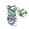
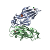
- PDB-6hm3: Crystal structure of Rad4 BRCT1,2 in complex with a Sld3 phosphop... -
+ Open data

- Basic information
| Entry | Database: PDB / ID: 6hm3 | ||||||
|---|---|---|---|---|---|---|---|
| Title | Crystal structure of Rad4 BRCT1,2 in complex with a Sld3 phosphopeptide | ||||||
 Components Components |
| ||||||
 Keywords Keywords | CELL CYCLE / BRCT domain phosphopeptide recognition | ||||||
| Function / homology |  Function and homology information Function and homology informationmitotic DNA damage checkpoint signaling / mitotic spindle pole body / DNA replication preinitiation complex / mitotic DNA replication checkpoint signaling / mitotic DNA replication initiation / mitotic G2 DNA damage checkpoint signaling / DNA replication initiation / signaling adaptor activity / meiotic cell cycle / mitotic spindle ...mitotic DNA damage checkpoint signaling / mitotic spindle pole body / DNA replication preinitiation complex / mitotic DNA replication checkpoint signaling / mitotic DNA replication initiation / mitotic G2 DNA damage checkpoint signaling / DNA replication initiation / signaling adaptor activity / meiotic cell cycle / mitotic spindle / site of double-strand break / chromatin / nucleus / cytosol Similarity search - Function | ||||||
| Biological species |  | ||||||
| Method |  X-RAY DIFFRACTION / SYNCHROTRON / MOLECULAR REPLACEMENT / Resolution: 1.77263620528 Å X-RAY DIFFRACTION / SYNCHROTRON / MOLECULAR REPLACEMENT / Resolution: 1.77263620528 Å | ||||||
 Authors Authors | Day, M. / Rappas, M. / Oliver, A.W. / Pearl, L.H. | ||||||
| Funding support |  United Kingdom, 1items United Kingdom, 1items
| ||||||
 Citation Citation | Journal: Elife / Year: 2018 Title: BRCT domains of the DNA damage checkpoint proteins TOPBP1/Rad4 display distinct specificities for phosphopeptide ligands. Authors: Day, M. / Rappas, M. / Ptasinska, K. / Boos, D. / Oliver, A.W. / Pearl, L.H. | ||||||
| History |
|
- Structure visualization
| Structure viewer | Molecule: MolmilJmol/JSmol |
|---|
- Downloads & links
-Download
| PDBx/mmCIF format | 6hm3.cif.gz | 75.7 KB | Display | PDBx/mmCIF format |
|---|---|---|---|---|
| PDB format | pdb6hm3.ent.gz | 44.4 KB | Display | PDB format |
| PDBx/mmJSON format | 6hm3.json.gz | Tree view | PDBx/mmJSON format | |
| Others |  Other downloads Other downloads |
-Validation report
| Arichive directory | https://data.pdbj.org/pub/pdb/validation_reports/hm/6hm3ftp://data.pdbj.org/pub/pdb/validation_reports/hm/6hm3 | HTTPS FTP |
|---|
-Related structure data
| Related structure data |  6hm4C  6hm5C  4bmcS S: Starting model for refinement C: citing same article ( |
|---|---|
| Similar structure data |


-Links
 PDBj
PDBj

- Assembly
| Deposited unit | 
| ||||||||||||
|---|---|---|---|---|---|---|---|---|---|---|---|---|---|
| 1 |
| ||||||||||||
| Unit cell |
|
-Components
| #1: Protein | Mass: 21236.457 Da / Num. of mol.: 1 Source method: isolated from a genetically manipulated source Source: (gene. exp.) Strain: 972 / ATCC 24843 / Gene: rad4, cut5, SPAC23C4.18c / Production host:  | ||||||
|---|---|---|---|---|---|---|---|
| #2: Protein/peptide | Mass: 3411.644 Da / Num. of mol.: 1 / Source method: obtained synthetically / Source: (synth.) | ||||||
| #3: Chemical |   Mass: 92.094 Da / Num. of mol.: 2 / Source method: obtained synthetically / Formula: C3H8O3 Mass: 92.094 Da / Num. of mol.: 2 / Source method: obtained synthetically / Formula: C3H8O3#4: Chemical |   Mass: 40.078 Da / Num. of mol.: 2 / Source method: obtained synthetically / Formula: Ca Mass: 40.078 Da / Num. of mol.: 2 / Source method: obtained synthetically / Formula: Ca#5: Water | ChemComp-HOH / |  Mass: 18.015 Da / Num. of mol.: 225 / Source method: isolated from a natural source / Formula: H2O Mass: 18.015 Da / Num. of mol.: 225 / Source method: isolated from a natural source / Formula: H2OHas protein modification | Y | |
-Experimental details
-Experiment
| Experiment | Method: X-RAY DIFFRACTION / Number of used crystals: 1 |
|---|
- Sample preparation
| Crystal | Density Matthews: 2.94 Å3/Da / Density % sol: 58.22 % |
|---|---|
| Crystal grow | Temperature: 287.15 K / Method: vapor diffusion, sitting drop / Details: Unknown |
-Data collection
| Diffraction | Mean temperature: 100 K |
|---|---|
| Diffraction source | Source: SYNCHROTRON / Site: Diamond / Beamline: I02 / Wavelength: 0.9795 Å |
| Detector | Type: ADSC QUANTUM 315 / Detector: CCD / Date: Oct 29, 2011 |
| Radiation | Protocol: SINGLE WAVELENGTH / Monochromatic (M) / Laue (L): M / Scattering type: x-ray |
| Radiation wavelength | Wavelength: 0.9795 Å / Relative weight: 1 |
| Reflection | Resolution: 1.77→42.2041207773 Å / Num. obs: 27589 / % possible obs: 98.3 % / Redundancy: 2.9 % / Biso Wilson estimate: 26.18 Å2 / Net I/σ(I): 15.7 |
| Reflection shell | Resolution: 1.77→1.82 Å |
- Processing
| Software |
| |||||||||||||||||||||||||||||||||||||||||||||||||||||||||||||||||||||||||||||
|---|---|---|---|---|---|---|---|---|---|---|---|---|---|---|---|---|---|---|---|---|---|---|---|---|---|---|---|---|---|---|---|---|---|---|---|---|---|---|---|---|---|---|---|---|---|---|---|---|---|---|---|---|---|---|---|---|---|---|---|---|---|---|---|---|---|---|---|---|---|---|---|---|---|---|---|---|---|---|
| Refinement | Method to determine structure: MOLECULAR REPLACEMENT Starting model: 4BMC Resolution: 1.77263620528→42.2041207773 Å / SU ML: 0.198039347248 / Cross valid method: FREE R-VALUE / σ(F): 1.3502924511 / Phase error: 24.8970812351 Stereochemistry target values: GeoStd + Monomer Library + CDL v1.2
| |||||||||||||||||||||||||||||||||||||||||||||||||||||||||||||||||||||||||||||
| Solvent computation | Shrinkage radii: 0.9 Å / VDW probe radii: 1.11 Å / Solvent model: FLAT BULK SOLVENT MODEL | |||||||||||||||||||||||||||||||||||||||||||||||||||||||||||||||||||||||||||||
| Displacement parameters | Biso mean: 34.5473330333 Å2 | |||||||||||||||||||||||||||||||||||||||||||||||||||||||||||||||||||||||||||||
| Refinement step | Cycle: LAST / Resolution: 1.77263620528→42.2041207773 Å
| |||||||||||||||||||||||||||||||||||||||||||||||||||||||||||||||||||||||||||||
| Refine LS restraints |
| |||||||||||||||||||||||||||||||||||||||||||||||||||||||||||||||||||||||||||||
| LS refinement shell |
|
