 Movie
Movie Controller

[English] 日本語
 Yorodumi
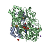
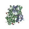
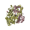
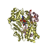
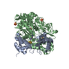
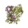
- PDB-6hli: wild-type NuoEF from Aquifex aeolicus - reduced form bound to NAD+ -
+ Open data

- Basic information
| Entry | Database: PDB / ID: 6hli | ||||||
|---|---|---|---|---|---|---|---|
| Title | wild-type NuoEF from Aquifex aeolicus - reduced form bound to NAD+ | ||||||
 Components Components | (NADH-quinone oxidoreductase subunit ...) x 2 | ||||||
 Keywords Keywords | ELECTRON TRANSPORT / Complex I / NuoEF / electron transfer / Aquifex aeolicus | ||||||
| Function / homology |  Function and homology information Function and homology informationTranslocases; Catalysing the translocation of protons; Linked to oxidoreductase reactions / NADH dehydrogenase (ubiquinone) activity / quinone binding / respiratory electron transport chain / 2 iron, 2 sulfur cluster binding / FMN binding / 4 iron, 4 sulfur cluster binding / oxidoreductase activity / metal ion binding Similarity search - Function | ||||||
| Biological species |   Aquifex aeolicus VF5 (bacteria) Aquifex aeolicus VF5 (bacteria) | ||||||
| Method |  X-RAY DIFFRACTION / MOLECULAR REPLACEMENT / Resolution: 2.38 Å X-RAY DIFFRACTION / MOLECULAR REPLACEMENT / Resolution: 2.38 Å | ||||||
 Authors Authors | Gerhardt, S. / Friedrich, T. / Einsle, O. / Gnandt, E. / Schulte, M. / Fiegen, D. | ||||||
| Funding support |  Germany, 1items Germany, 1items
| ||||||
 Citation Citation | Journal: Nat Commun / Year: 2019 Title: A mechanism to prevent production of reactive oxygen species by Escherichia coli respiratory complex I. Authors: Schulte, M. / Frick, K. / Gnandt, E. / Jurkovic, S. / Burschel, S. / Labatzke, R. / Aierstock, K. / Fiegen, D. / Wohlwend, D. / Gerhardt, S. / Einsle, O. / Friedrich, T. | ||||||
| History |
|
- Structure visualization
| Structure viewer | Molecule: MolmilJmol/JSmol |
|---|
- Downloads & links
-Download
| PDBx/mmCIF format | 6hli.cif.gz | 479.1 KB | Display | PDBx/mmCIF format |
|---|---|---|---|---|
| PDB format | pdb6hli.ent.gz | 393 KB | Display | PDB format |
| PDBx/mmJSON format | 6hli.json.gz | Tree view | PDBx/mmJSON format | |
| Others |  Other downloads Other downloads |
-Validation report
| Arichive directory | https://data.pdbj.org/pub/pdb/validation_reports/hl/6hliftp://data.pdbj.org/pub/pdb/validation_reports/hl/6hli | HTTPS FTP |
|---|
-Related structure data
| Related structure data |  6hl2C  6hl3C  6hl4C  6hlaC  6hljC  6hlmC  6q9cC  6q9gC  6q9jC 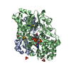 6q9kC  6r7pC C: citing same article ( |
|---|---|
| Similar structure data |


-Links
 PDBj
PDBj


- Assembly
| Deposited unit | 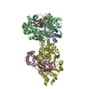
| ||||||||
|---|---|---|---|---|---|---|---|---|---|
| 1 | 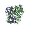
| ||||||||
| 2 | 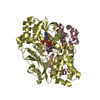
| ||||||||
| Unit cell |
|
-Components
-NADH-quinone oxidoreductase subunit ... , 2 types, 4 molecules ACBD
| #1: Protein | Mass: 18573.619 Da / Num. of mol.: 2 Source method: isolated from a genetically manipulated source Source: (gene. exp.) Aquifex aeolicus VF5 (bacteria) / Gene: nuoE, aq_574 / Production host: #2: Protein | Mass: 48523.301 Da / Num. of mol.: 2 Source method: isolated from a genetically manipulated source Details: Sequence contains C-terminal 6xHis Expressions-tag / Source: (gene. exp.) Aquifex aeolicus VF5 (bacteria) / Gene: nuoF, aq_573 / Production host: |
|---|
-Non-polymers , 5 types, 256 molecules 


| #3: Chemical |  Mass: 175.820 Da / Num. of mol.: 2 / Source method: obtained synthetically / Formula: Fe2S2 Mass: 175.820 Da / Num. of mol.: 2 / Source method: obtained synthetically / Formula: Fe2S2#4: Chemical |  Mass: 351.640 Da / Num. of mol.: 2 / Source method: obtained synthetically / Formula: Fe4S4 Mass: 351.640 Da / Num. of mol.: 2 / Source method: obtained synthetically / Formula: Fe4S4#5: Chemical |  Mass: 456.344 Da / Num. of mol.: 2 / Source method: obtained synthetically / Formula: C17H21N4O9P Mass: 456.344 Da / Num. of mol.: 2 / Source method: obtained synthetically / Formula: C17H21N4O9P#6: Chemical |  Mass: 663.425 Da / Num. of mol.: 2 / Source method: obtained synthetically / Formula: C21H27N7O14P2 / Comment: NAD*YM Mass: 663.425 Da / Num. of mol.: 2 / Source method: obtained synthetically / Formula: C21H27N7O14P2 / Comment: NAD*YM#7: Water | ChemComp-HOH / | Mass: 18.015 Da / Num. of mol.: 248 / Source method: isolated from a natural source / Formula: H2O |
|---|
-Experimental details
-Experiment
| Experiment | Method: X-RAY DIFFRACTION / Number of used crystals: 1 |
|---|
- Sample preparation
| Crystal | Density Matthews: 2.61 Å3/Da / Density % sol: 52.88 % |
|---|---|
| Crystal grow | Temperature: 293 K / Method: vapor diffusion, sitting drop / pH: 6.9 / Details: 0.1M Tris/HCl 0.1M Sodium citrate / PH range: 6.9 - 7.1 |
-Data collection
| Diffraction | Mean temperature: 100 K |
|---|---|
| Diffraction source | Source: ROTATING ANODE / Type: RIGAKU MICROMAX-007 / Wavelength: 1.54179 Å |
| Detector | Type: MARRESEARCH / Detector: IMAGE PLATE / Date: Jul 22, 2016 / Details: VARIMAX |
| Radiation | Protocol: SINGLE WAVELENGTH / Monochromatic (M) / Laue (L): M / Scattering type: x-ray |
| Radiation wavelength | Wavelength: 1.54179 Å / Relative weight: 1 |
| Reflection | Resolution: 2.38→116 Å / Num. obs: 57523 / % possible obs: 100 % / Redundancy: 6.4 % / Biso Wilson estimate: 45.9 Å2 / CC1/2: 0.998 / Rmerge(I) obs: 0.119 / Rpim(I) all: 0.051 / Rsym value: 0.119 / Net I/σ(I): 13.2 |
| Reflection shell | Resolution: 2.38→2.51 Å / Redundancy: 6.4 % / Rmerge(I) obs: 0.757 / Mean I/σ(I) obs: 2.2 / Num. unique obs: 8284 / CC1/2: 0.778 / Rpim(I) all: 0.351 / Rsym value: 0.757 / % possible all: 100 |
- Processing
| Software |
| |||||||||||||||||||||||||||||||||||||||||||||||||||||||||||||||||||||||||||||||||||||||||||||||||||||||||||||||||||||||||||||
|---|---|---|---|---|---|---|---|---|---|---|---|---|---|---|---|---|---|---|---|---|---|---|---|---|---|---|---|---|---|---|---|---|---|---|---|---|---|---|---|---|---|---|---|---|---|---|---|---|---|---|---|---|---|---|---|---|---|---|---|---|---|---|---|---|---|---|---|---|---|---|---|---|---|---|---|---|---|---|---|---|---|---|---|---|---|---|---|---|---|---|---|---|---|---|---|---|---|---|---|---|---|---|---|---|---|---|---|---|---|---|---|---|---|---|---|---|---|---|---|---|---|---|---|---|---|---|
| Refinement | Method to determine structure: MOLECULAR REPLACEMENT Starting model: IN-HOUSE Resolution: 2.38→60.3 Å / Cor.coef. Fo:Fc: 0.919 / Cor.coef. Fo:Fc free: 0.903 / SU R Cruickshank DPI: 0.343 / Cross valid method: THROUGHOUT / σ(F): 0 / SU R Blow DPI: 0.345 / SU Rfree Blow DPI: 0.228 / SU Rfree Cruickshank DPI: 0.23
| |||||||||||||||||||||||||||||||||||||||||||||||||||||||||||||||||||||||||||||||||||||||||||||||||||||||||||||||||||||||||||||
| Displacement parameters | Biso mean: 50.48 Å2
| |||||||||||||||||||||||||||||||||||||||||||||||||||||||||||||||||||||||||||||||||||||||||||||||||||||||||||||||||||||||||||||
| Refinement step | Cycle: 1 / Resolution: 2.38→60.3 Å
| |||||||||||||||||||||||||||||||||||||||||||||||||||||||||||||||||||||||||||||||||||||||||||||||||||||||||||||||||||||||||||||
| Refine LS restraints |
| |||||||||||||||||||||||||||||||||||||||||||||||||||||||||||||||||||||||||||||||||||||||||||||||||||||||||||||||||||||||||||||
| LS refinement shell | Resolution: 2.38→2.44 Å / Total num. of bins used: 20
| |||||||||||||||||||||||||||||||||||||||||||||||||||||||||||||||||||||||||||||||||||||||||||||||||||||||||||||||||||||||||||||
| Refinement TLS params. | Method: refined / Refine-ID: X-RAY DIFFRACTION
| |||||||||||||||||||||||||||||||||||||||||||||||||||||||||||||||||||||||||||||||||||||||||||||||||||||||||||||||||||||||||||||
| Refinement TLS group |
|
