 Movie
Movie Controller


+ Open data

- Basic information
| Entry | Database: PDB / ID: 6hfw | ||||||
|---|---|---|---|---|---|---|---|
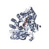
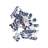
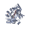
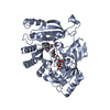
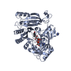
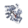
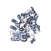
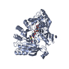
| Title | Mycobacterium tuberculosis DprE1 in complex with CMP1 | ||||||
 Components Components | Decaprenylphosphoryl-beta-D-ribose oxidase | ||||||
 Keywords Keywords | OXIDOREDUCTASE / OXIDOREDUCTASE INHIBITOR | ||||||
| Function / homology |  Function and homology information Function and homology informationarabinan biosynthetic process / cell wall polysaccharide biosynthetic process / decaprenylphospho-beta-D-ribofuranose 2-dehydrogenase / D-arabinono-1,4-lactone oxidase activity / capsule polysaccharide biosynthetic process / FAD binding / cell wall organization / periplasmic space / oxidoreductase activity / response to antibiotic / plasma membrane Similarity search - Function | ||||||
| Biological species |   Mycobacterium tuberculosis (bacteria) Mycobacterium tuberculosis (bacteria) | ||||||
| Method |  X-RAY DIFFRACTION / SYNCHROTRON / Resolution: 2.47 Å X-RAY DIFFRACTION / SYNCHROTRON / Resolution: 2.47 Å | ||||||
 Authors Authors | Chung, C. | ||||||
 Citation Citation | Journal: Sci Rep / Year: 2018 Title: Novel insight into the reaction of nitro, nitroso and hydroxylamino benzothiazinones and of benzoxacinones with Mycobacterium tuberculosis DprE1. Authors: Richter, A. / Rudolph, I. / Mollmann, U. / Voigt, K. / Chung, C.W. / Singh, O.M.P. / Rees, M. / Mendoza-Losana, A. / Bates, R. / Ballell, L. / Batt, S. / Veerapen, N. / Futterer, K. / Besra, ...Authors: Richter, A. / Rudolph, I. / Mollmann, U. / Voigt, K. / Chung, C.W. / Singh, O.M.P. / Rees, M. / Mendoza-Losana, A. / Bates, R. / Ballell, L. / Batt, S. / Veerapen, N. / Futterer, K. / Besra, G. / Imming, P. / Argyrou, A. | ||||||
| History |
|
- Structure visualization
| Structure viewer | Molecule: MolmilJmol/JSmol |
|---|
- Downloads & links
-Download
| PDBx/mmCIF format | 6hfw.cif.gz | 352.5 KB | Display | PDBx/mmCIF format |
|---|---|---|---|---|
| PDB format | pdb6hfw.ent.gz | 285.9 KB | Display | PDB format |
| PDBx/mmJSON format | 6hfw.json.gz | Tree view | PDBx/mmJSON format | |
| Others |  Other downloads Other downloads |
-Validation report
| Arichive directory | https://data.pdbj.org/pub/pdb/validation_reports/hf/6hfwftp://data.pdbj.org/pub/pdb/validation_reports/hf/6hfw | HTTPS FTP |
|---|
-Related structure data


-Links
 PDBj
PDBj


- Assembly
| Deposited unit | 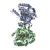
| ||||||||
|---|---|---|---|---|---|---|---|---|---|
| 1 | 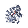
| ||||||||
| 2 | 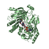
| ||||||||
| Unit cell |
|
-Components
| #1: Protein | Mass: 51838.664 Da / Num. of mol.: 2 Source method: isolated from a genetically manipulated source Source: (gene. exp.) Mycobacterium tuberculosis (strain ATCC 25618 / H37Rv) (bacteria)Strain: ATCC 25618 / H37Rv / Gene: dprE1, Rv3790 / Production host: References: UniProt: P9WJF1, decaprenylphospho-beta-D-ribofuranose 2-dehydrogenase #2: Chemical |   Mass: 785.550 Da / Num. of mol.: 2 / Source method: obtained synthetically / Formula: C27H33N9O15P2 / Comment: FAD*YM Mass: 785.550 Da / Num. of mol.: 2 / Source method: obtained synthetically / Formula: C27H33N9O15P2 / Comment: FAD*YM#3: Chemical | ChemComp-IMD / |   Mass: 69.085 Da / Num. of mol.: 1 / Source method: obtained synthetically / Formula: C3H5N2 Mass: 69.085 Da / Num. of mol.: 1 / Source method: obtained synthetically / Formula: C3H5N2#4: Chemical |   Mass: 345.340 Da / Num. of mol.: 2 / Source method: obtained synthetically / Formula: C14H14F3N3O2S Mass: 345.340 Da / Num. of mol.: 2 / Source method: obtained synthetically / Formula: C14H14F3N3O2S#5: Water | ChemComp-HOH / |  Mass: 18.015 Da / Num. of mol.: 228 / Source method: isolated from a natural source / Formula: H2O Mass: 18.015 Da / Num. of mol.: 228 / Source method: isolated from a natural source / Formula: H2O |
|---|
-Experimental details
-Experiment
| Experiment | Method: X-RAY DIFFRACTION / Number of used crystals: 1 |
|---|
- Sample preparation
| Crystal | Density Matthews: 2.52 Å3/Da / Density % sol: 51.22 % |
|---|---|
| Crystal grow | Temperature: 298 K / Method: microbatch / Details: 20mM in 30% PPG, 100mM Imidazole,pH 7.4 |
-Data collection
| Diffraction | Mean temperature: 100 K |
|---|---|
| Diffraction source | Source: SYNCHROTRON / Site: ESRF  / Beamline: ID29 / Wavelength: 1.0725 Å / Beamline: ID29 / Wavelength: 1.0725 Å |
| Detector | Type: DECTRIS PILATUS3 6M / Detector: PIXEL / Date: Sep 25, 2013 |
| Radiation | Protocol: SINGLE WAVELENGTH / Monochromatic (M) / Laue (L): M / Scattering type: x-ray |
| Radiation wavelength | Wavelength: 1.0725 Å / Relative weight: 1 |
| Reflection | Resolution: 2.47→56.69 Å / Num. obs: 33896 / % possible obs: 96.8 % / Redundancy: 2.9 % / Net I/σ(I): 11.3 |
| Reflection shell | Resolution: 2.47→2.6 Å |
- Processing
| Software |
| ||||||||||||||||||||||||||||||||||||||||||||||||||||||||||||||||||||||||||||||||||||||||||||||||||||||||||||||||||||||||||||||||||||||||||||||||||||||||||||||||||||||||||||||||||||||
|---|---|---|---|---|---|---|---|---|---|---|---|---|---|---|---|---|---|---|---|---|---|---|---|---|---|---|---|---|---|---|---|---|---|---|---|---|---|---|---|---|---|---|---|---|---|---|---|---|---|---|---|---|---|---|---|---|---|---|---|---|---|---|---|---|---|---|---|---|---|---|---|---|---|---|---|---|---|---|---|---|---|---|---|---|---|---|---|---|---|---|---|---|---|---|---|---|---|---|---|---|---|---|---|---|---|---|---|---|---|---|---|---|---|---|---|---|---|---|---|---|---|---|---|---|---|---|---|---|---|---|---|---|---|---|---|---|---|---|---|---|---|---|---|---|---|---|---|---|---|---|---|---|---|---|---|---|---|---|---|---|---|---|---|---|---|---|---|---|---|---|---|---|---|---|---|---|---|---|---|---|---|---|---|
| Refinement | Resolution: 2.47→56.69 Å / Cor.coef. Fo:Fc: 0.966 / Cor.coef. Fo:Fc free: 0.942 / SU B: 15.939 / SU ML: 0.173 / Cross valid method: THROUGHOUT / ESU R: 0.441 / ESU R Free: 0.252 / Details: HYDROGENS HAVE BEEN ADDED IN THE RIDING POSITIONS
| ||||||||||||||||||||||||||||||||||||||||||||||||||||||||||||||||||||||||||||||||||||||||||||||||||||||||||||||||||||||||||||||||||||||||||||||||||||||||||||||||||||||||||||||||||||||
| Solvent computation | Ion probe radii: 0.8 Å / Shrinkage radii: 0.8 Å / VDW probe radii: 1.2 Å | ||||||||||||||||||||||||||||||||||||||||||||||||||||||||||||||||||||||||||||||||||||||||||||||||||||||||||||||||||||||||||||||||||||||||||||||||||||||||||||||||||||||||||||||||||||||
| Displacement parameters | Biso mean: 55.09 Å2
| ||||||||||||||||||||||||||||||||||||||||||||||||||||||||||||||||||||||||||||||||||||||||||||||||||||||||||||||||||||||||||||||||||||||||||||||||||||||||||||||||||||||||||||||||||||||
| Refinement step | Cycle: 1 / Resolution: 2.47→56.69 Å
| ||||||||||||||||||||||||||||||||||||||||||||||||||||||||||||||||||||||||||||||||||||||||||||||||||||||||||||||||||||||||||||||||||||||||||||||||||||||||||||||||||||||||||||||||||||||
| Refine LS restraints |
|