 Movie
Movie Controller


+ Open data

- Basic information
| Entry | Database: PDB / ID: 6hfg | ||||||
|---|---|---|---|---|---|---|---|
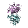
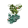
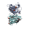
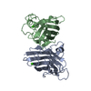
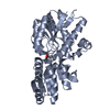
| Title | Structure of the REC114 PH domain | ||||||
 Components Components | Meiotic recombination protein REC114 | ||||||
 Keywords Keywords | RECOMBINATION / Pleckstrin Homology domain Meiotic recombination | ||||||
| Function / homology | Meiotic recombination protein REC114-like / Meiotic recombination protein REC114-like / meiotic DNA double-strand break formation / oogenesis / chromosome / DNA recombination / spermatogenesis / Meiotic recombination protein REC114 Function and homology information Function and homology information | ||||||
| Biological species |  | ||||||
| Method |  X-RAY DIFFRACTION / SYNCHROTRON / SAD / Resolution: 2.5 Å X-RAY DIFFRACTION / SYNCHROTRON / SAD / Resolution: 2.5 Å | ||||||
 Authors Authors | Juarez-Martinez, A.B. / de Massy, B. / Kadlec, J. | ||||||
 Citation Citation | Journal: Life Sci Alliance / Year: 2018 Title: Mouse REC114 is essential for meiotic DNA double-strand break formation and forms a complex with MEI4. Authors: Kumar, R. / Oliver, C. / Brun, C. / Juarez-Martinez, A.B. / Tarabay, Y. / Kadlec, J. / de Massy, B. | ||||||
| History |
|
- Structure visualization
| Structure viewer | Molecule: MolmilJmol/JSmol |
|---|
- Downloads & links
-Download
| PDBx/mmCIF format | 6hfg.cif.gz | 36.7 KB | Display | PDBx/mmCIF format |
|---|---|---|---|---|
| PDB format | pdb6hfg.ent.gz | 24 KB | Display | PDB format |
| PDBx/mmJSON format | 6hfg.json.gz | Tree view | PDBx/mmJSON format | |
| Others |  Other downloads Other downloads |
-Validation report
| Arichive directory | https://data.pdbj.org/pub/pdb/validation_reports/hf/6hfgftp://data.pdbj.org/pub/pdb/validation_reports/hf/6hfg | HTTPS FTP |
|---|
-Related structure data
| Similar structure data |
|---|


-Links
 PDBj
PDBj- Assembly
| Deposited unit | 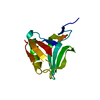
| ||||||||
|---|---|---|---|---|---|---|---|---|---|
| 1 | 
| ||||||||
| Unit cell |
|
-Components
| #1: Protein | Mass: 16239.387 Da / Num. of mol.: 1 Source method: isolated from a genetically manipulated source Source: (gene. exp.)  |
|---|
-Experimental details
-Experiment
| Experiment | Method: X-RAY DIFFRACTION / Number of used crystals: 1 |
|---|
- Sample preparation
| Crystal | Density Matthews: 4.25 Å3/Da / Density % sol: 71.07 % |
|---|---|
| Crystal grow | Temperature: 293.15 K / Method: vapor diffusion, sitting drop / pH: 6.5 Details: 0.25 M ammonium sulphate, 0.1 M MES (pH 6.5), 28% PEG 5000 MME |
-Data collection
| Diffraction | Mean temperature: 100 K |
|---|---|
| Diffraction source | Source: SYNCHROTRON / Site: ESRF  / Beamline: MASSIF-1 / Wavelength: 0.966 Å / Beamline: MASSIF-1 / Wavelength: 0.966 Å |
| Detector | Type: DECTRIS PILATUS3 2M / Detector: PIXEL / Date: Oct 29, 2017 |
| Radiation | Protocol: SINGLE WAVELENGTH / Monochromatic (M) / Laue (L): M / Scattering type: x-ray |
| Radiation wavelength | Wavelength: 0.966 Å / Relative weight: 1 |
| Reflection | Resolution: 2.5→93 Å / Num. obs: 10262 / % possible obs: 99.6 % / Observed criterion σ(I): 1.1 / Redundancy: 11.2 % / Rmerge(I) obs: 0.057 / Net I/σ(I): 24.2 |
| Reflection shell | Resolution: 2.5→2.57 Å / Redundancy: 11.1 % / Mean I/σ(I) obs: 1.1 / Num. unique obs: 749 / CC1/2: 0.548 / % possible all: 97.5 |
- Processing
| Software |
| ||||||||||||||||||||||||||||||||||||||||||||||||||||||||||||||||||||||||||||||||||||||||||||||||||||||||||||||||||||||||||||||||||||||||||||||||||||||||||||||||||||||||||||||||||||||
|---|---|---|---|---|---|---|---|---|---|---|---|---|---|---|---|---|---|---|---|---|---|---|---|---|---|---|---|---|---|---|---|---|---|---|---|---|---|---|---|---|---|---|---|---|---|---|---|---|---|---|---|---|---|---|---|---|---|---|---|---|---|---|---|---|---|---|---|---|---|---|---|---|---|---|---|---|---|---|---|---|---|---|---|---|---|---|---|---|---|---|---|---|---|---|---|---|---|---|---|---|---|---|---|---|---|---|---|---|---|---|---|---|---|---|---|---|---|---|---|---|---|---|---|---|---|---|---|---|---|---|---|---|---|---|---|---|---|---|---|---|---|---|---|---|---|---|---|---|---|---|---|---|---|---|---|---|---|---|---|---|---|---|---|---|---|---|---|---|---|---|---|---|---|---|---|---|---|---|---|---|---|---|---|
| Refinement | Method to determine structure: SAD / Resolution: 2.5→46.59 Å / Cor.coef. Fo:Fc: 0.941 / Cor.coef. Fo:Fc free: 0.902 / SU B: 8.425 / SU ML: 0.178 / Cross valid method: THROUGHOUT / ESU R: 0.235 / ESU R Free: 0.225 / Details: HYDROGENS HAVE BEEN ADDED IN THE RIDING POSITIONS
| ||||||||||||||||||||||||||||||||||||||||||||||||||||||||||||||||||||||||||||||||||||||||||||||||||||||||||||||||||||||||||||||||||||||||||||||||||||||||||||||||||||||||||||||||||||||
| Solvent computation | Ion probe radii: 0.8 Å / Shrinkage radii: 0.8 Å / VDW probe radii: 1.2 Å | ||||||||||||||||||||||||||||||||||||||||||||||||||||||||||||||||||||||||||||||||||||||||||||||||||||||||||||||||||||||||||||||||||||||||||||||||||||||||||||||||||||||||||||||||||||||
| Displacement parameters | Biso mean: 77.071 Å2
| ||||||||||||||||||||||||||||||||||||||||||||||||||||||||||||||||||||||||||||||||||||||||||||||||||||||||||||||||||||||||||||||||||||||||||||||||||||||||||||||||||||||||||||||||||||||
| Refinement step | Cycle: 1 / Resolution: 2.5→46.59 Å
| ||||||||||||||||||||||||||||||||||||||||||||||||||||||||||||||||||||||||||||||||||||||||||||||||||||||||||||||||||||||||||||||||||||||||||||||||||||||||||||||||||||||||||||||||||||||
| Refine LS restraints |
|