Evidence: gel filtration, Ligand binding characterised FRET and SPR
Type
Name
Symmetry operation
Number
identity operation
1_555
x,y,z
1
Buried area
790 Å2
ΔGint
-19 kcal/mol
Surface area
8310 Å2
Method
PISA
Unit cell
Length a, b, c (Å)
79.070, 79.070, 138.388
Angle α, β, γ (deg.)
90.00, 90.00, 120.00
Int Tables number
179
Space group name H-M
P6522
Components on special symmetry positions
ID
Model
Components
1
1
A-1203-
SO4
2
1
A-1203-
SO4
3
1
A-1413-
HOH
-
Components
#1: Protein
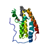
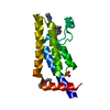
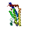
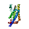
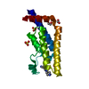
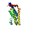
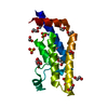
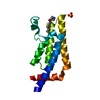
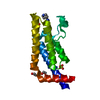
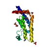
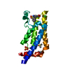
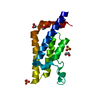
ATPasefamilyAAAdomain-containingprotein2 / AAA nuclear coregulator cancer-associated protein / ANCCA
Mass: 15453.514 Da / Num. of mol.: 1 Source method: isolated from a genetically manipulated source Details: SM remains from tag cleavage / Source: (gene. exp.) Homo sapiens (human) / Gene: ATAD2, L16, PRO2000 / Plasmid: pNIC28-Bsa4 / Production host: Escherichia coli (E. coli) / References: UniProt: Q6PL18, EC: 3.6.1.3
Protocol: SINGLE WAVELENGTH / Monochromatic (M) / Laue (L): M / Scattering type: x-ray
Radiation wavelength
Wavelength: 1.54178 Å / Relative weight: 1
Reflection
Resolution: 1.9→68.48 Å / Num. obs: 20781 / % possible obs: 99.6 % / Redundancy: 11.28 % / Net I/σ(I): 12.3
Reflection shell
Resolution: 1.9→1.97 Å
-
Processing
Software
Name
Version
Classification
REFMAC
5.8.0073
refinement
d*TREK
datareduction
d*TREK
datascaling
Refinement
Resolution: 1.9→68.48 Å / Cor.coef. Fo:Fc: 0.954 / Cor.coef. Fo:Fc free: 0.927 / SU B: 5.15 / SU ML: 0.082 / Cross valid method: THROUGHOUT / ESU R: 0.115 / ESU R Free: 0.116 / Details: HYDROGENS HAVE BEEN ADDED IN THE RIDING POSITIONS
Rfactor
Num. reflection
% reflection
Selection details
Rfree
0.22956
1066
5.1 %
RANDOM
Rwork
0.19378
-
-
-
obs
0.19561
19709
99.55 %
-
Solvent computation
Ion probe radii: 0.8 Å / Shrinkage radii: 0.8 Å / VDW probe radii: 1.2 Å
 Movie
Movie Controller

 Yorodumi
 Open data
 Basic information
 Components
 Keywords
 Function and homology information
 Homo sapiens (human)
 X-RAY DIFFRACTION / Resolution: 1.9 Å
  Authors
 Citation
 Structure visualization
 Downloads & links
 Other downloads

 PDBj
PDBj


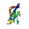
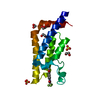
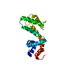
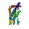
 Assembly
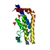


 Mass: 96.063 Da / Num. of mol.: 3 / Source method: obtained synthetically / Formula: SO4
 Mass: 62.068 Da / Num. of mol.: 2 / Source method: obtained synthetically / Formula: C2H6O2
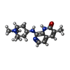
 Mass: 298.383 Da / Num. of mol.: 1 / Source method: obtained synthetically / Formula: C17H22N4O
 Mass: 18.015 Da / Num. of mol.: 198 / Source method: isolated from a natural source / Formula: H2O
 Sample preparation
 Processing