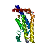
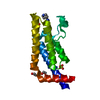
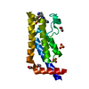
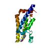
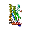
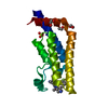
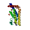
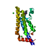
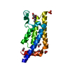
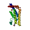
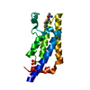
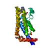
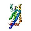
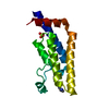
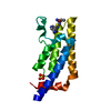
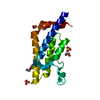
Entry Database : PDB / ID : 5a5pTitle Crystal structure of human ATAD2 bromodomain in complex with 8-2-(dimethylamino)ethylamino-3-methyl-1,2-dihydroquinolin-2-one ATPASE FAMILY AAA DOMAIN-CONTAINING PROTEIN 2 Keywords / / / / / Function / homology Function Domain/homology Component
/ / / / / / / / / / / / / / / / / / / / / / / / / / / / / / / / / / / / / / / / / / / / / / / / Biological species HOMO SAPIENS (human)Method / / Resolution : 2.03 Å Authors Chung, C. / Bamborough, P. / Demont, E. Journal : J.Med.Chem. / Year : 2015Title : Fragment-Based Discovery of Low-Micromolar Atad2 Bromodomain Inhibitors.Authors: Demont, E.H. / Chung, C. / Furze, R.C. / Grandi, P. / Michon, A. / Wellaway, C. / Barrett, N. / Bridges, A.M. / Craggs, P.D. / Diallo, H. / Dixon, D.P. / Douault, C. / Emmons, A.J. / Jones, ... Authors : Demont, E.H. / Chung, C. / Furze, R.C. / Grandi, P. / Michon, A. / Wellaway, C. / Barrett, N. / Bridges, A.M. / Craggs, P.D. / Diallo, H. / Dixon, D.P. / Douault, C. / Emmons, A.J. / Jones, E.J. / Karamshi, B.V. / Locke, K. / Mitchell, D.J. / Mouzon, B.H. / Prinjha, R.K. / Roberts, A.D. / Sheppard, R.J. / Watson, R.J. / Bamborough, P. History Deposition Jun 20, 2015 Deposition site / Processing site Revision 1.0 Jul 22, 2015 Provider / Type Revision 1.1 Aug 5, 2015 Group Revision 1.2 May 8, 2024 Group Data collection / Database references ... Data collection / Database references / Derived calculations / Other Category chem_comp_atom / chem_comp_bond ... chem_comp_atom / chem_comp_bond / database_2 / pdbx_database_status / struct_site Item _database_2.pdbx_DOI / _database_2.pdbx_database_accession ... _database_2.pdbx_DOI / _database_2.pdbx_database_accession / _pdbx_database_status.status_code_sf / _struct_site.pdbx_auth_asym_id / _struct_site.pdbx_auth_comp_id / _struct_site.pdbx_auth_seq_id
Show all Show less
 Movie
Movie Controller

 Yorodumi
 Open data
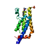
 Basic information
 Components
 Keywords
 Function and homology information
 HOMO SAPIENS (human)
 X-RAY DIFFRACTION /
  Authors
 Citation
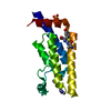
 Structure visualization
 Downloads & links
 Other downloads
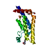


 PDBj
PDBj


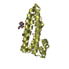
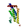
 Assembly
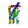


 Mass: 62.068 Da / Num. of mol.: 2 / Source method: obtained synthetically / Formula: C2H6O2
 Mass: 245.320 Da / Num. of mol.: 1 / Source method: obtained synthetically / Formula: C14H19N3O
 Mass: 96.063 Da / Num. of mol.: 1 / Source method: obtained synthetically / Formula: SO4
 Mass: 18.015 Da / Num. of mol.: 215 / Source method: isolated from a natural source / Formula: H2O
 Sample preparation
 Processing