 Movie
Movie Controller


+ Open data

- Basic information
| Entry | Database: PDB / ID: 6ha4 | |||||||||
|---|---|---|---|---|---|---|---|---|---|---|
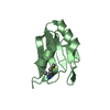
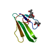
| Title | Crystal structure of PAF - p-sulfonatocalix[4]arene complex | |||||||||
 Components Components | Pc24g00380 protein | |||||||||
 Keywords Keywords | ANTIFUNGAL PROTEIN / Penicillium chrysogenum / calixarene / molecular glues / nucleating agent | |||||||||
| Function / homology |  Function and homology information Function and homology information | |||||||||
| Biological species |  Penicillium rubens (fungus) Penicillium rubens (fungus) | |||||||||
| Method |  X-RAY DIFFRACTION / SYNCHROTRON / MOLECULAR REPLACEMENT / Resolution: 1.33 Å X-RAY DIFFRACTION / SYNCHROTRON / MOLECULAR REPLACEMENT / Resolution: 1.33 Å | |||||||||
 Authors Authors | Alex, J.M. / Rennie, M. / Engilberge, S. / Batta, G. / Crowley, P.B. | |||||||||
| Funding support |  Ireland, 2items Ireland, 2items
| |||||||||
 Citation Citation | Journal: Iucrj / Year: 2019 Title: Calixarene-mediated assembly of a small antifungal protein. Authors: Alex, J.M. / Rennie, M.L. / Engilberge, S. / Lehoczki, G. / Dorottya, H. / Fizil, A. / Batta, G. / Crowley, P.B. | |||||||||
| History |
|
- Structure visualization
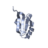
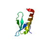
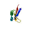
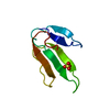
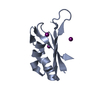
| Structure viewer | Molecule: MolmilJmol/JSmol |
|---|
- Downloads & links
-Download
| PDBx/mmCIF format | 6ha4.cif.gz | 38.9 KB | Display | PDBx/mmCIF format |
|---|---|---|---|---|
| PDB format | pdb6ha4.ent.gz | 25.8 KB | Display | PDB format |
| PDBx/mmJSON format | 6ha4.json.gz | Tree view | PDBx/mmJSON format | |
| Others |  Other downloads Other downloads |
-Validation report
| Arichive directory | https://data.pdbj.org/pub/pdb/validation_reports/ha/6ha4ftp://data.pdbj.org/pub/pdb/validation_reports/ha/6ha4 | HTTPS FTP |
|---|
-Related structure data
| Related structure data | 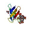 6hahC 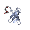 6hajC 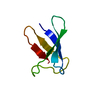 2mhvS S: Starting model for refinement C: citing same article ( |
|---|---|
| Similar structure data |


-Links
 PDBj
PDBj
- Assembly
| Deposited unit | 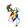
| ||||||||
|---|---|---|---|---|---|---|---|---|---|
| 1 |
| ||||||||
| Unit cell |
|
-Components
| #1: Protein | Mass: 6263.099 Da / Num. of mol.: 1 / Source method: isolated from a natural source Source: (natural) Penicillium rubens (strain ATCC 28089 / DSM 1075 / NRRL 1951 / Wisconsin 54-1255) (fungus)References: UniProt: B6HWK0 |
|---|---|
| #2: Chemical | ChemComp-GOL /   Mass: 92.094 Da / Num. of mol.: 1 / Source method: obtained synthetically / Formula: C3H8O3 Mass: 92.094 Da / Num. of mol.: 1 / Source method: obtained synthetically / Formula: C3H8O3 |
| #3: Chemical | ChemComp-T3Y / 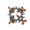  Mass: 744.741 Da / Num. of mol.: 1 / Source method: obtained synthetically / Formula: C28H24O16S4 / Feature type: SUBJECT OF INVESTIGATION Mass: 744.741 Da / Num. of mol.: 1 / Source method: obtained synthetically / Formula: C28H24O16S4 / Feature type: SUBJECT OF INVESTIGATION |
| #4: Water | ChemComp-HOH /  Mass: 18.015 Da / Num. of mol.: 55 / Source method: isolated from a natural source / Formula: H2O Mass: 18.015 Da / Num. of mol.: 55 / Source method: isolated from a natural source / Formula: H2O |
| Has protein modification | Y |
-Experimental details
-Experiment
| Experiment | Method: X-RAY DIFFRACTION / Number of used crystals: 1 |
|---|
- Sample preparation
| Crystal | Density Matthews: 1.9 Å3/Da / Density % sol: 35.15 % |
|---|---|
| Crystal grow | Temperature: 293 K / Method: vapor diffusion, hanging drop / pH: 5.6 / Details: 30% PEG 3350 + 0.05 M Sodium acetate pH 5.6 |
-Data collection
| Diffraction | Mean temperature: 100 K |
|---|---|
| Diffraction source | Source: SYNCHROTRON / Site: SOLEIL  / Beamline: PROXIMA 2 / Wavelength: 0.98 Å / Beamline: PROXIMA 2 / Wavelength: 0.98 Å |
| Detector | Type: DECTRIS EIGER X 9M / Detector: PIXEL / Date: Sep 15, 2016 |
| Radiation | Protocol: SINGLE WAVELENGTH / Monochromatic (M) / Laue (L): M / Scattering type: x-ray |
| Radiation wavelength | Wavelength: 0.98 Å / Relative weight: 1 |
| Reflection | Resolution: 1.3→27.8 Å / Num. obs: 10142 / % possible obs: 92.2 % / Redundancy: 3.4 % / Biso Wilson estimate: 16.37 Å2 / CC1/2: 0.98 / Rpim(I) all: 0.062 / Rrim(I) all: 0.012 / Net I/av σ(I): 6.1 / Net I/σ(I): 6.1 |
| Reflection shell | Resolution: 1.33→1.37 Å / Mean I/σ(I) obs: 1.3 / CC1/2: 0.34 / Rpim(I) all: 0.34 / Rrim(I) all: 0.61 |
- Processing
| Software |
| ||||||||||||||||||||||||||||||||||||||||||||||||||||||||||||||||||||||||||||||||||||||||||||||||||||||||||||||||||
|---|---|---|---|---|---|---|---|---|---|---|---|---|---|---|---|---|---|---|---|---|---|---|---|---|---|---|---|---|---|---|---|---|---|---|---|---|---|---|---|---|---|---|---|---|---|---|---|---|---|---|---|---|---|---|---|---|---|---|---|---|---|---|---|---|---|---|---|---|---|---|---|---|---|---|---|---|---|---|---|---|---|---|---|---|---|---|---|---|---|---|---|---|---|---|---|---|---|---|---|---|---|---|---|---|---|---|---|---|---|---|---|---|---|---|---|
| Refinement | Method to determine structure: MOLECULAR REPLACEMENT Starting model: 2MHV Resolution: 1.33→27.8 Å / Cor.coef. Fo:Fc: 0.953 / Cor.coef. Fo:Fc free: 0.911 / SU R Cruickshank DPI: 0.076 / Cross valid method: THROUGHOUT / σ(F): 0 / SU R Blow DPI: 0.071 / SU Rfree Blow DPI: 0.072 / SU Rfree Cruickshank DPI: 0.069
| ||||||||||||||||||||||||||||||||||||||||||||||||||||||||||||||||||||||||||||||||||||||||||||||||||||||||||||||||||
| Displacement parameters | Biso mean: 20.96 Å2
| ||||||||||||||||||||||||||||||||||||||||||||||||||||||||||||||||||||||||||||||||||||||||||||||||||||||||||||||||||
| Refine analyze | Luzzati coordinate error obs: 0.18 Å | ||||||||||||||||||||||||||||||||||||||||||||||||||||||||||||||||||||||||||||||||||||||||||||||||||||||||||||||||||
| Refinement step | Cycle: 1 / Resolution: 1.33→27.8 Å
| ||||||||||||||||||||||||||||||||||||||||||||||||||||||||||||||||||||||||||||||||||||||||||||||||||||||||||||||||||
| Refine LS restraints |
| ||||||||||||||||||||||||||||||||||||||||||||||||||||||||||||||||||||||||||||||||||||||||||||||||||||||||||||||||||
| LS refinement shell | Resolution: 1.33→1.48 Å / Total num. of bins used: 5
| ||||||||||||||||||||||||||||||||||||||||||||||||||||||||||||||||||||||||||||||||||||||||||||||||||||||||||||||||||
| Refinement TLS params. | Method: refined / Origin x: -3.7966 Å / Origin y: 4.5228 Å / Origin z: 31.3802 Å
| ||||||||||||||||||||||||||||||||||||||||||||||||||||||||||||||||||||||||||||||||||||||||||||||||||||||||||||||||||
| Refinement TLS group | Selection details: { A|* } |
