 Movie
Movie Controller

[English] 日本語
 Yorodumi
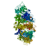
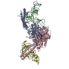
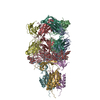
- PDB-6h6g: Crystal Structure of TcdB2-TccC3 without hypervariable C-terminal... -
+ Open data

- Basic information
| Entry | Database: PDB / ID: 6h6g | ||||||
|---|---|---|---|---|---|---|---|
| Title | Crystal Structure of TcdB2-TccC3 without hypervariable C-terminal region | ||||||
 Components Components | TcdB2,TccC3 | ||||||
 Keywords Keywords | TOXIN / Tc toxin / ABC toxin | ||||||
| Function / homology |  Function and homology information Function and homology information | ||||||
| Biological species |  Photorhabdus luminescens (bacteria) Photorhabdus luminescens (bacteria) | ||||||
| Method |  X-RAY DIFFRACTION / SYNCHROTRON / MOLECULAR REPLACEMENT / Resolution: 3.004 Å X-RAY DIFFRACTION / SYNCHROTRON / MOLECULAR REPLACEMENT / Resolution: 3.004 Å | ||||||
 Authors Authors | Gatsogiannis, C. / Merino, F. / Roderer, D. / Balchin, D. / Schubert, E. / Kuhlee, A. / Hayer-Hartl, M. / Raunser, S. | ||||||
| Funding support |  Germany, 1items Germany, 1items
| ||||||
 Citation Citation | Journal: Nature / Year: 2018 Title: Tc toxin activation requires unfolding and refolding of a β-propeller. Authors: Christos Gatsogiannis / Felipe Merino / Daniel Roderer / David Balchin / Evelyn Schubert / Anne Kuhlee / Manajit Hayer-Hartl / Stefan Raunser / Abstract: Tc toxins secrete toxic enzymes into host cells using a unique syringe-like injection mechanism. They are composed of three subunits, TcA, TcB and TcC. TcA forms the translocation channel and the TcB- ...Tc toxins secrete toxic enzymes into host cells using a unique syringe-like injection mechanism. They are composed of three subunits, TcA, TcB and TcC. TcA forms the translocation channel and the TcB-TcC heterodimer functions as a cocoon that shields the toxic enzyme. Binding of the cocoon to the channel triggers opening of the cocoon and translocation of the toxic enzyme into the channel. Here we show in atomic detail how the assembly of the three components activates the toxin. We find that part of the cocoon completely unfolds and refolds into an alternative conformation upon binding. The presence of the toxic enzyme inside the cocoon is essential for its subnanomolar binding affinity for the TcA subunit. The enzyme passes through a narrow negatively charged constriction site inside the cocoon, probably acting as an extruder that releases the unfolded protein with its C terminus first into the translocation channel. | ||||||
| History |
|
- Structure visualization
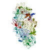
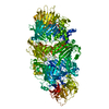
| Structure viewer | Molecule: MolmilJmol/JSmol |
|---|
- Downloads & links
-Download
| PDBx/mmCIF format | 6h6g.cif.gz | 787.3 KB | Display | PDBx/mmCIF format |
|---|---|---|---|---|
| PDB format | pdb6h6g.ent.gz | 653.9 KB | Display | PDB format |
| PDBx/mmJSON format | 6h6g.json.gz | Tree view | PDBx/mmJSON format | |
| Others |  Other downloads Other downloads |
-Validation report
| Arichive directory | https://data.pdbj.org/pub/pdb/validation_reports/h6/6h6gftp://data.pdbj.org/pub/pdb/validation_reports/h6/6h6g | HTTPS FTP |
|---|
-Related structure data
| Related structure data |  0149C  0150C 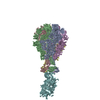 6h6eC 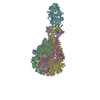 6h6fC  4o9xS S: Starting model for refinement C: citing same article ( |
|---|---|
| Similar structure data |

-Links
 PDBj
PDBj
- Assembly
| Deposited unit | 
| ||||||||
|---|---|---|---|---|---|---|---|---|---|
| 1 |
| ||||||||
| Unit cell |
|
-Components
| #1: Protein | Mass: 243770.109 Da / Num. of mol.: 1 Source method: isolated from a genetically manipulated source Source: (gene. exp.) Photorhabdus luminescens (bacteria) / Gene: tcdB2, TccC3 / Production host: |
|---|---|
| #2: Water | ChemComp-HOH /  Mass: 18.015 Da / Num. of mol.: 135 / Source method: isolated from a natural source / Formula: H2O Mass: 18.015 Da / Num. of mol.: 135 / Source method: isolated from a natural source / Formula: H2O |
-Experimental details
-Experiment
| Experiment | Method: X-RAY DIFFRACTION / Number of used crystals: 1 |
|---|
- Sample preparation
| Crystal | Density Matthews: 4.5 Å3/Da / Density % sol: 72.66 % |
|---|---|
| Crystal grow | Temperature: 293.15 K / Method: vapor diffusion, sitting drop Details: 0.1 M tri-sodium citrate pH 5.5, 10 % PEG 8000, 10 % ethylenglycol |
-Data collection
| Diffraction | Mean temperature: 100 K |
|---|---|
| Diffraction source | Source: SYNCHROTRON / Site: SLS  / Beamline: X10SA / Wavelength: 0.97794 Å / Beamline: X10SA / Wavelength: 0.97794 Å |
| Detector | Type: PSI PILATUS 6M / Detector: PIXEL / Date: Feb 6, 2017 |
| Radiation | Protocol: SINGLE WAVELENGTH / Monochromatic (M) / Laue (L): M / Scattering type: x-ray |
| Radiation wavelength | Wavelength: 0.97794 Å / Relative weight: 1 |
| Reflection | Resolution: 3→50 Å / Num. obs: 87208 / % possible obs: 99.9 % / Redundancy: 20 % / Net I/σ(I): 7.3 |
| Reflection shell | Resolution: 3→3.08 Å / Rmerge(I) obs: 0.5751 / Rpim(I) all: 0.129 / Rrim(I) all: 0.5895 |
- Processing
| Software |
| |||||||||||||||||||||||||||||||||||||||||||||||||||||||||||||||||||||||||||||||||||||||||||||||||||||||||||||||||||||||||||||||||||||||||||||||||||||||||||||||||||||||||||||||||||||||||||||||||||||||||||||||||||||||||
|---|---|---|---|---|---|---|---|---|---|---|---|---|---|---|---|---|---|---|---|---|---|---|---|---|---|---|---|---|---|---|---|---|---|---|---|---|---|---|---|---|---|---|---|---|---|---|---|---|---|---|---|---|---|---|---|---|---|---|---|---|---|---|---|---|---|---|---|---|---|---|---|---|---|---|---|---|---|---|---|---|---|---|---|---|---|---|---|---|---|---|---|---|---|---|---|---|---|---|---|---|---|---|---|---|---|---|---|---|---|---|---|---|---|---|---|---|---|---|---|---|---|---|---|---|---|---|---|---|---|---|---|---|---|---|---|---|---|---|---|---|---|---|---|---|---|---|---|---|---|---|---|---|---|---|---|---|---|---|---|---|---|---|---|---|---|---|---|---|---|---|---|---|---|---|---|---|---|---|---|---|---|---|---|---|---|---|---|---|---|---|---|---|---|---|---|---|---|---|---|---|---|---|---|---|---|---|---|---|---|---|---|---|---|---|---|---|---|---|
| Refinement | Method to determine structure: MOLECULAR REPLACEMENT Starting model: 4O9X Resolution: 3.004→48.639 Å / SU ML: 0.43 / Cross valid method: FREE R-VALUE / σ(F): 1.33 / Phase error: 27.33 / Stereochemistry target values: ML
| |||||||||||||||||||||||||||||||||||||||||||||||||||||||||||||||||||||||||||||||||||||||||||||||||||||||||||||||||||||||||||||||||||||||||||||||||||||||||||||||||||||||||||||||||||||||||||||||||||||||||||||||||||||||||
| Solvent computation | Shrinkage radii: 0.9 Å / VDW probe radii: 1.11 Å / Solvent model: FLAT BULK SOLVENT MODEL | |||||||||||||||||||||||||||||||||||||||||||||||||||||||||||||||||||||||||||||||||||||||||||||||||||||||||||||||||||||||||||||||||||||||||||||||||||||||||||||||||||||||||||||||||||||||||||||||||||||||||||||||||||||||||
| Refinement step | Cycle: LAST / Resolution: 3.004→48.639 Å
| |||||||||||||||||||||||||||||||||||||||||||||||||||||||||||||||||||||||||||||||||||||||||||||||||||||||||||||||||||||||||||||||||||||||||||||||||||||||||||||||||||||||||||||||||||||||||||||||||||||||||||||||||||||||||
| Refine LS restraints |
| |||||||||||||||||||||||||||||||||||||||||||||||||||||||||||||||||||||||||||||||||||||||||||||||||||||||||||||||||||||||||||||||||||||||||||||||||||||||||||||||||||||||||||||||||||||||||||||||||||||||||||||||||||||||||
| LS refinement shell |
|
