Movie
Movie Controller

[English] 日本語
 Yorodumi
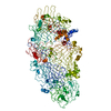
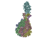
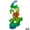
- PDB-6h6e: PTC3 holotoxin complex from Photorhabdus luminecens in prepore st... -
+ Open data

- Basic information
| Entry | Database: PDB / ID: 6h6e | |||||||||
|---|---|---|---|---|---|---|---|---|---|---|
| Title | PTC3 holotoxin complex from Photorhabdus luminecens in prepore state (TcdA1, TcdB2, TccC3) | |||||||||
 Components Components |
| |||||||||
 Keywords Keywords | TOXIN / pore forming toxin / translocation / bacterial toxin / a-PFT / Photorhabdus / ABC | |||||||||
| Function / homology |  Function and homology information Function and homology information | |||||||||
| Biological species |  Photorhabdus luminescens (bacteria) Photorhabdus luminescens (bacteria) | |||||||||
| Method | ELECTRON MICROSCOPY / single particle reconstruction / cryo EM / Resolution: 3.95 Å | |||||||||
 Authors Authors | Gatsogiannis, C. / Merino, F. / Roderer, D. / Balchin, D. / Schubert, E. / Kuhlee, A. / Hayer-Hartl, M. / Raunser, S. | |||||||||
| Funding support |  Germany, 2items Germany, 2items
| |||||||||
 Citation Citation | Journal: Nature / Year: 2018 Title: Tc toxin activation requires unfolding and refolding of a β-propeller. Authors: Christos Gatsogiannis / Felipe Merino / Daniel Roderer / David Balchin / Evelyn Schubert / Anne Kuhlee / Manajit Hayer-Hartl / Stefan Raunser / Abstract: Tc toxins secrete toxic enzymes into host cells using a unique syringe-like injection mechanism. They are composed of three subunits, TcA, TcB and TcC. TcA forms the translocation channel and the TcB- ...Tc toxins secrete toxic enzymes into host cells using a unique syringe-like injection mechanism. They are composed of three subunits, TcA, TcB and TcC. TcA forms the translocation channel and the TcB-TcC heterodimer functions as a cocoon that shields the toxic enzyme. Binding of the cocoon to the channel triggers opening of the cocoon and translocation of the toxic enzyme into the channel. Here we show in atomic detail how the assembly of the three components activates the toxin. We find that part of the cocoon completely unfolds and refolds into an alternative conformation upon binding. The presence of the toxic enzyme inside the cocoon is essential for its subnanomolar binding affinity for the TcA subunit. The enzyme passes through a narrow negatively charged constriction site inside the cocoon, probably acting as an extruder that releases the unfolded protein with its C terminus first into the translocation channel. | |||||||||
| History |
|
- Structure visualization
| Movie |
Movie viewer |
|---|---|
| Structure viewer | Molecule: MolmilJmol/JSmol |
- Downloads & links
-Download
| PDBx/mmCIF format | 6h6e.cif.gz | 2.6 MB | Display | PDBx/mmCIF format |
|---|---|---|---|---|
| PDB format | pdb6h6e.ent.gz | Display | PDB format | |
| PDBx/mmJSON format | 6h6e.json.gz | Tree view | PDBx/mmJSON format | |
| Others |  Other downloads Other downloads |
-Validation report
| Arichive directory | https://data.pdbj.org/pub/pdb/validation_reports/h6/6h6eftp://data.pdbj.org/pub/pdb/validation_reports/h6/6h6e | HTTPS FTP |
|---|
-Related structure data
| Related structure data |  0149MC  0150C  6h6fC  6h6gC M: map data used to model this data C: citing same article ( |
|---|---|
| Similar structure data |

-Links
 PDBj
PDBj
- Assembly
| Deposited unit | 
|
|---|---|
| 1 |
|
-Components
| #1: Protein | Mass: 283229.406 Da / Num. of mol.: 5 Source method: isolated from a genetically manipulated source Source: (gene. exp.) Photorhabdus luminescens (bacteria) / Gene: tcdA, tcdA1 / Production host: #2: Protein | | Mass: 273678.656 Da / Num. of mol.: 1 Source method: isolated from a genetically manipulated source Source: (gene. exp.) Photorhabdus luminescens (bacteria) / Gene: tcdB2, TccC3 / Production host: |
|---|
-Experimental details
-Experiment
| Experiment | Method: ELECTRON MICROSCOPY |
|---|---|
| EM experiment | Aggregation state: PARTICLE / 3D reconstruction method: single particle reconstruction |
- Sample preparation
| Component |
| ||||||||||||||||||||||||||||
|---|---|---|---|---|---|---|---|---|---|---|---|---|---|---|---|---|---|---|---|---|---|---|---|---|---|---|---|---|---|
| Molecular weight |
| ||||||||||||||||||||||||||||
| Source (natural) |
| ||||||||||||||||||||||||||||
| Source (recombinant) |
| ||||||||||||||||||||||||||||
| Buffer solution | pH: 8 | ||||||||||||||||||||||||||||
| Buffer component |
| ||||||||||||||||||||||||||||
| Specimen | Embedding applied: NO / Shadowing applied: NO / Staining applied: NO / Vitrification applied: YES | ||||||||||||||||||||||||||||
| Specimen support | Grid material: COPPER / Grid mesh size: 400 divisions/in. / Grid type: C-flat-2/1 | ||||||||||||||||||||||||||||
| Vitrification | Instrument: GATAN CRYOPLUNGE 3 / Cryogen name: ETHANE / Humidity: 94 % / Details: blot 3 sec before plunging |
- Electron microscopy imaging
| Experimental equipment |  Model: Titan Krios / Image courtesy: FEI Company |
|---|---|
| Microscopy | Model: FEI TITAN KRIOS |
| Electron gun | Electron source:  FIELD EMISSION GUN / Accelerating voltage: 300 kV / Illumination mode: OTHER FIELD EMISSION GUN / Accelerating voltage: 300 kV / Illumination mode: OTHER |
| Electron lens | Mode: BRIGHT FIELD |
| Specimen holder | Cryogen: NITROGEN / Specimen holder model: FEI TITAN KRIOS AUTOGRID HOLDER |
| Image recording | Electron dose: 2.2 e/Å2 / Film or detector model: FEI FALCON II (4k x 4k) / Num. of real images: 3068 |
- Processing
| EM software |
| ||||||||||||||||||||||||||||||||||||||||||||||||||||||||||||
|---|---|---|---|---|---|---|---|---|---|---|---|---|---|---|---|---|---|---|---|---|---|---|---|---|---|---|---|---|---|---|---|---|---|---|---|---|---|---|---|---|---|---|---|---|---|---|---|---|---|---|---|---|---|---|---|---|---|---|---|---|---|
| CTF correction | Type: PHASE FLIPPING AND AMPLITUDE CORRECTION | ||||||||||||||||||||||||||||||||||||||||||||||||||||||||||||
| Particle selection | Num. of particles selected: 205336 | ||||||||||||||||||||||||||||||||||||||||||||||||||||||||||||
| Symmetry | Point symmetry: C1 (asymmetric) | ||||||||||||||||||||||||||||||||||||||||||||||||||||||||||||
| 3D reconstruction | Resolution: 3.95 Å / Resolution method: FSC 0.143 CUT-OFF / Num. of particles: 89148 / Symmetry type: POINT | ||||||||||||||||||||||||||||||||||||||||||||||||||||||||||||
| Atomic model building | B value: 68.03 / Protocol: OTHER / Space: REAL / Target criteria: Model to Map FSC | ||||||||||||||||||||||||||||||||||||||||||||||||||||||||||||
| Atomic model building | 3D fitting-ID: 1 / Source name: PDB / Type: experimental model
|