 Movie
Movie Controller

[English] 日本語
 Yorodumi
- PDB-6h37: The crystal structure of human carbonic anhydrase VII in complex ... -
+ Open data

- Basic information
| Entry | Database: PDB / ID: 6h37 | ||||||
|---|---|---|---|---|---|---|---|
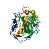
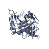
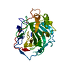
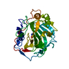
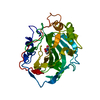
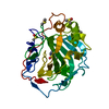
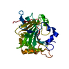
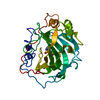
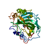
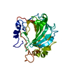
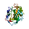
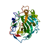
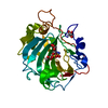
| Title | The crystal structure of human carbonic anhydrase VII in complex with 4-(4-phenyl)-4-hydroxy-1-piperidine-1-carbonyl)benzenesulfonamide | ||||||
 Components Components | Carbonic anhydrase 7 | ||||||
 Keywords Keywords | LYASE / Protein-inhibitor binding | ||||||
| Function / homology |  Function and homology information Function and homology information: / regulation of chloride transport / Reversible hydration of carbon dioxide / positive regulation of synaptic transmission, GABAergic / carbonic anhydrase / regulation of intracellular pH / carbonate dehydratase activity / neuron cellular homeostasis / zinc ion binding / cytosol / cytoplasm Similarity search - Function | ||||||
| Biological species |  Homo sapiens (human) Homo sapiens (human) | ||||||
| Method |  X-RAY DIFFRACTION / FOURIER SYNTHESIS / Resolution: 1.9 Å X-RAY DIFFRACTION / FOURIER SYNTHESIS / Resolution: 1.9 Å | ||||||
 Authors Authors | Buemi, M.R. / Di Fiore, A. / De Luca, L. / Ferro, S. / Mancuso, F. / Monti, S.M. / Buonanno, M. / Angeli, A. / Russo, E. / De Sarro, G. ...Buemi, M.R. / Di Fiore, A. / De Luca, L. / Ferro, S. / Mancuso, F. / Monti, S.M. / Buonanno, M. / Angeli, A. / Russo, E. / De Sarro, G. / Supuran, C.T. / De Simone, G. / Gitto, R. | ||||||
 Citation Citation | Journal: Eur J Med Chem / Year: 2018 Title: Exploring structural properties of potent human carbonic anhydrase inhibitors bearing a 4-(cycloalkylamino-1-carbonyl)benzenesulfonamide moiety. Authors: Buemi, M.R. / Di Fiore, A. / De Luca, L. / Angeli, A. / Mancuso, F. / Ferro, S. / Monti, S.M. / Buonanno, M. / Russo, E. / De Sarro, G. / De Simone, G. / Supuran, C.T. / Gitto, R. | ||||||
| History |
|
- Structure visualization
| Structure viewer | Molecule: MolmilJmol/JSmol |
|---|
- Downloads & links
-Download
| PDBx/mmCIF format | 6h37.cif.gz | 69.7 KB | Display | PDBx/mmCIF format |
|---|---|---|---|---|
| PDB format | pdb6h37.ent.gz | 49.2 KB | Display | PDB format |
| PDBx/mmJSON format | 6h37.json.gz | Tree view | PDBx/mmJSON format | |
| Others |  Other downloads Other downloads |
-Validation report
| Arichive directory | https://data.pdbj.org/pub/pdb/validation_reports/h3/6h37ftp://data.pdbj.org/pub/pdb/validation_reports/h3/6h37 | HTTPS FTP |
|---|
-Related structure data
| Related structure data | 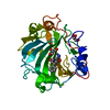 6h2zC 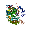 6h33C 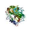 6h34C 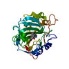 6h36C  6h38C  6g4tS C: citing same article ( S: Starting model for refinement |
|---|---|
| Similar structure data |


-Links
 PDBj
PDBj- Assembly
| Deposited unit | 
| ||||||||
|---|---|---|---|---|---|---|---|---|---|
| 1 |
| ||||||||
| Unit cell |
|
-Components
| #1: Protein | Mass: 30925.693 Da / Num. of mol.: 1 / Mutation: C183S, C217S Source method: isolated from a genetically manipulated source Source: (gene. exp.) Homo sapiens (human) / Gene: CA7 / Production host:  |
|---|---|
| #2: Chemical | ChemComp-ZN /   Mass: 65.409 Da / Num. of mol.: 1 / Source method: obtained synthetically / Formula: Zn Mass: 65.409 Da / Num. of mol.: 1 / Source method: obtained synthetically / Formula: Zn |
| #3: Chemical | ChemComp-FKQ /   Mass: 360.427 Da / Num. of mol.: 1 / Source method: obtained synthetically / Formula: C18H20N2O4S Mass: 360.427 Da / Num. of mol.: 1 / Source method: obtained synthetically / Formula: C18H20N2O4S |
| #4: Water | ChemComp-HOH /  Mass: 18.015 Da / Num. of mol.: 115 / Source method: isolated from a natural source / Formula: H2O Mass: 18.015 Da / Num. of mol.: 115 / Source method: isolated from a natural source / Formula: H2O |
| Has protein modification | Y |
-Experimental details
-Experiment
| Experiment | Method: X-RAY DIFFRACTION / Number of used crystals: 1 |
|---|
- Sample preparation
| Crystal | Density Matthews: 2.09 Å3/Da / Density % sol: 41.07 % |
|---|---|
| Crystal grow | Temperature: 293 K / Method: vapor diffusion, hanging drop / pH: 8.5 Details: 25% v/v Peg3350, 0.2 M Ammonium acetate, 0.1 M Tris pH 8.5 |
-Data collection
| Diffraction | Mean temperature: 100 K |
|---|---|
| Diffraction source | Source: ROTATING ANODE / Type: RIGAKU MICROMAX-007 HF / Wavelength: 1.54178 Å |
| Detector | Type: RIGAKU SATURN 944 / Detector: CCD / Date: Feb 26, 2018 / Details: Mirror |
| Radiation | Monochromator: SI 111 CHANNEL / Protocol: SINGLE WAVELENGTH / Monochromatic (M) / Laue (L): M / Scattering type: x-ray |
| Radiation wavelength | Wavelength: 1.54178 Å / Relative weight: 1 |
| Reflection | Resolution: 1.9→30.9 Å / Num. obs: 69317 / % possible obs: 92.1 % / Redundancy: 3.6 % / Rmerge(I) obs: 0.046 / Rpim(I) all: 0.024 / Rrim(I) all: 0.052 / Net I/σ(I): 22.3 |
| Reflection shell | Resolution: 1.9→1.97 Å / Redundancy: 2.9 % / Rmerge(I) obs: 0.181 / Mean I/σ(I) obs: 7.4 / Rpim(I) all: 0.108 / Rrim(I) all: 0.213 / % possible all: 69.4 |
- Processing
| Software |
| ||||||||||||||||
|---|---|---|---|---|---|---|---|---|---|---|---|---|---|---|---|---|---|
| Refinement | Method to determine structure: FOURIER SYNTHESIS Starting model: 6G4T Resolution: 1.9→30.9 Å / Cross valid method: FREE R-VALUE
| ||||||||||||||||
| Refinement step | Cycle: LAST / Resolution: 1.9→30.9 Å
|
