 Movie
Movie Controller

[English] 日本語
 Yorodumi
- PDB-3t5z: Crystal structure of the human carbonic anhydrase II in complex w... -
+ Open data

- Basic information
| Entry | Database: PDB / ID: 3t5z | ||||||
|---|---|---|---|---|---|---|---|
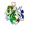
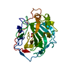
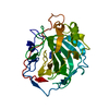
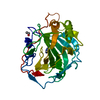
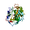
| Title | Crystal structure of the human carbonic anhydrase II in complex with N-methoxy-benzenesulfonamide | ||||||
 Components Components | Carbonic anhydrase 2 | ||||||
 Keywords Keywords | LYASE/LYASE INHIBITOR / Protein-inhibitor complex / LYASE-LYASE INHIBITOR complex | ||||||
| Function / homology |  Function and homology information Function and homology informationpositive regulation of dipeptide transmembrane transport / : / secretion / regulation of monoatomic anion transport / cyanamide hydratase / cyanamide hydratase activity / arylesterase activity / regulation of chloride transport / Reversible hydration of carbon dioxide / positive regulation of synaptic transmission, GABAergic ...positive regulation of dipeptide transmembrane transport / : / secretion / regulation of monoatomic anion transport / cyanamide hydratase / cyanamide hydratase activity / arylesterase activity / regulation of chloride transport / Reversible hydration of carbon dioxide / positive regulation of synaptic transmission, GABAergic / morphogenesis of an epithelium / angiotensin-activated signaling pathway / Developmental Lineage of Pancreatic Ductal Cells / carbonic anhydrase / regulation of intracellular pH / carbonate dehydratase activity / carbon dioxide transport / neuron cellular homeostasis / Erythrocytes take up oxygen and release carbon dioxide / Erythrocytes take up carbon dioxide and release oxygen / apical part of cell / myelin sheath / extracellular exosome / zinc ion binding / plasma membrane / cytosol / cytoplasm Similarity search - Function | ||||||
| Biological species |  Homo sapiens (human) Homo sapiens (human) | ||||||
| Method |  X-RAY DIFFRACTION / FOURIER SYNTHESIS / Resolution: 1.65 Å X-RAY DIFFRACTION / FOURIER SYNTHESIS / Resolution: 1.65 Å | ||||||
 Authors Authors | Di Fiore, A. / Maresca, A. / Alterio, V. / Supuran, C.T. / De Simone, G. | ||||||
 Citation Citation | Journal: Chem.Commun.(Camb.) / Year: 2011 Title: Carbonic anhydrase inhibitors: X-ray crystallographic studies for the binding of N-substituted benzenesulfonamides to human isoform II. Authors: Di Fiore, A. / Maresca, A. / Alterio, V. / Supuran, C.T. / De Simone, G. #1: Journal: Proteins / Year: 1988Title: Refined structure of human carbonic anhydrase II at 2.0 A resolution. Authors: Eriksson, A.E. / Jones, T.A. / Liljas, A. #2: Journal: Chemmedchem / Year: 2009Title: Thermodynamic optimisation in drug discovery: a case study using carbonic anhydrase inhibitors. Authors: Scott, A.D. / Phillips, C. / Alex, A. / Flocco, M. / Bent, A. / Randall, A. / O'Brien, R. / Damian, L. / Jones, L.H. | ||||||
| History |
|
- Structure visualization
| Structure viewer | Molecule: MolmilJmol/JSmol |
|---|
- Downloads & links
-Download
| PDBx/mmCIF format | 3t5z.cif.gz | 75.2 KB | Display | PDBx/mmCIF format |
|---|---|---|---|---|
| PDB format | pdb3t5z.ent.gz | 54.1 KB | Display | PDB format |
| PDBx/mmJSON format | 3t5z.json.gz | Tree view | PDBx/mmJSON format | |
| Others |  Other downloads Other downloads |
-Validation report
| Arichive directory | https://data.pdbj.org/pub/pdb/validation_reports/t5/3t5zftp://data.pdbj.org/pub/pdb/validation_reports/t5/3t5z | HTTPS FTP |
|---|
-Related structure data
| Related structure data | 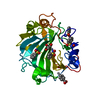 3t5uC 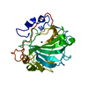 1ca2S S: Starting model for refinement C: citing same article ( |
|---|---|
| Similar structure data |


-Links
 PDBj
PDBj

- Assembly
| Deposited unit | 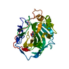
| ||||||||
|---|---|---|---|---|---|---|---|---|---|
| 1 |
| ||||||||
| Unit cell |
|
-Components
| #1: Protein | Mass: 29157.863 Da / Num. of mol.: 1 / Source method: isolated from a natural source / Source: (natural) Homo sapiens (human) / References: UniProt: P00918, carbonic anhydrase |
|---|---|
| #2: Chemical | ChemComp-ZN /   Mass: 65.409 Da / Num. of mol.: 1 / Source method: obtained synthetically / Formula: Zn Mass: 65.409 Da / Num. of mol.: 1 / Source method: obtained synthetically / Formula: Zn |
| #3: Chemical | ChemComp-B09 /   Mass: 187.216 Da / Num. of mol.: 1 / Source method: obtained synthetically / Formula: C7H9NO3S Mass: 187.216 Da / Num. of mol.: 1 / Source method: obtained synthetically / Formula: C7H9NO3S |
| #4: Chemical | ChemComp-MBO /   Mass: 321.703 Da / Num. of mol.: 1 / Source method: obtained synthetically / Formula: C7H5HgO2 Mass: 321.703 Da / Num. of mol.: 1 / Source method: obtained synthetically / Formula: C7H5HgO2 |
| #5: Water | ChemComp-HOH /  Mass: 18.015 Da / Num. of mol.: 356 / Source method: isolated from a natural source / Formula: H2O Mass: 18.015 Da / Num. of mol.: 356 / Source method: isolated from a natural source / Formula: H2O |
-Experimental details
-Experiment
| Experiment | Method: X-RAY DIFFRACTION / Number of used crystals: 1 |
|---|
- Sample preparation
| Crystal | Density Matthews: 2.08 Å3/Da / Density % sol: 40.78 % |
|---|---|
| Crystal grow | Temperature: 293 K / Method: vapor diffusion, hanging drop / pH: 8.5 Details: 2.6 M ammonium sulfate, 0.3 M sodium chloride, 0.1 M Tris-HCl, pH 8.5, 5 mM 4-hydroxymercurybenzoate, VAPOR DIFFUSION, HANGING DROP, temperature 293K |
-Data collection
| Diffraction | Mean temperature: 100 K |
|---|---|
| Diffraction source | Source: ROTATING ANODE / Type: RIGAKU / Wavelength: 1.5418 Å |
| Detector | Type: RIGAKU SATURN / Detector: CCD / Date: Feb 8, 2011 |
| Radiation | Protocol: SINGLE WAVELENGTH / Monochromatic (M) / Laue (L): M / Scattering type: x-ray |
| Radiation wavelength | Wavelength: 1.5418 Å / Relative weight: 1 |
| Reflection | Resolution: 1.65→20 Å / Num. all: 28042 / Num. obs: 28042 / % possible obs: 96.4 % / Redundancy: 2.8 % / Rmerge(I) obs: 0.043 / Net I/σ(I): 21.03 |
| Reflection shell | Resolution: 1.65→1.71 Å / Rmerge(I) obs: 0.061 / Mean I/σ(I) obs: 13.45 / Num. unique all: 2511 / % possible all: 87.6 |
- Processing
| Software |
| |||||||||||||||||||||||||
|---|---|---|---|---|---|---|---|---|---|---|---|---|---|---|---|---|---|---|---|---|---|---|---|---|---|---|
| Refinement | Method to determine structure: FOURIER SYNTHESIS Starting model: PDB ENTRY 1CA2 Resolution: 1.65→20 Å / Occupancy max: 1 / Occupancy min: 0.5 / σ(F): 0 / Stereochemistry target values: Engh & Huber
| |||||||||||||||||||||||||
| Solvent computation | Bsol: 43.3279 Å2 | |||||||||||||||||||||||||
| Displacement parameters | Biso max: 52.48 Å2 / Biso mean: 13.6681 Å2 / Biso min: 2.34 Å2
| |||||||||||||||||||||||||
| Refinement step | Cycle: LAST / Resolution: 1.65→20 Å
| |||||||||||||||||||||||||
| Refine LS restraints |
| |||||||||||||||||||||||||
| Xplor file |
|
