| Entry | Database: PDB / ID: 6h2f
|
|---|
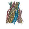
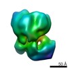
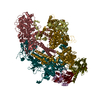
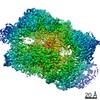
| Title | Structure of the pre-pore AhlB of the tripartite alpha-pore forming toxin, AHL, from Aeromonas hydrophila. |
|---|
 Components Components | AhlB |
|---|
 Keywords Keywords | TOXIN / Tripartite pore-forming toxin |
|---|
| Function / homology |  Function and homology information Function and homology information
Hemolysin BL-binding component / Bacillus haemolytic enterotoxin (HBL) / : / Hemolysin E; Chain: A; / Hemolysin E; Chain: A; - #10 / Up-down Bundle / Mainly AlphaSimilarity search - Domain/homology |
|---|
| Biological species |  Aeromonas hydrophila AL09-71 (bacteria) Aeromonas hydrophila AL09-71 (bacteria) |
|---|
| Method |  X-RAY DIFFRACTION / SYNCHROTRON / MOLECULAR REPLACEMENT / Resolution: 2.55 Å X-RAY DIFFRACTION / SYNCHROTRON / MOLECULAR REPLACEMENT / Resolution: 2.55 Å |
|---|
 Authors Authors | Churchill-Angus, A.M. / Wilson, J.S. / Baker, P.J. |
|---|
| Funding support |  United Kingdom, 1items United Kingdom, 1items | Organization | Grant number | Country |
|---|
| Biotechnology and Biological Sciences Research Council | BB/F016832/1 | United Kingdom |
|
|---|
 Citation Citation | Journal: Nat Commun / Year: 2019
Title: Identification and structural analysis of the tripartite alpha-pore forming toxin of Aeromonas hydrophila.
Authors: Wilson, J.S. / Churchill-Angus, A.M. / Davies, S.P. / Sedelnikova, S.E. / Tzokov, S.B. / Rafferty, J.B. / Bullough, P.A. / Bisson, C. / Baker, P.J. |
|---|
| History | | Deposition | Jul 13, 2018 | Deposition site: PDBE / Processing site: PDBE |
|---|
| Revision 1.0 | Jul 10, 2019 | Provider: repository / Type: Initial release |
|---|
| Revision 1.1 | Jul 17, 2019 | Group: Data collection / Database references / Category: citation / citation_author
Item: _citation.journal_volume / _citation.page_first ..._citation.journal_volume / _citation.page_first / _citation.page_last / _citation.pdbx_database_id_PubMed / _citation.title |
|---|
| Revision 1.2 | May 1, 2024 | Group: Data collection / Database references / Refinement description
Category: chem_comp_atom / chem_comp_bond ...chem_comp_atom / chem_comp_bond / database_2 / pdbx_initial_refinement_model
Item: _database_2.pdbx_DOI / _database_2.pdbx_database_accession |
|---|
|
|---|
 Movie
Movie Controller

 Yorodumi
 Open data
 Basic information
 Structure visualization
 Downloads & links
 Other downloads


 PDBj
PDBj Assembly
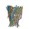

 Mass: 94.971 Da / Num. of mol.: 4 / Source method: obtained synthetically / Formula: PO4
 Sample preparation
 Processing