 Movie
Movie Controller

[English] 日本語
 Yorodumi
- EMDB-2808: Electron cryo-microscopy of the HerA-NurA double strand break res... -
+ Open data

- Basic information
| Entry | Database: EMDB / ID: EMD-2808 | |||||||||
|---|---|---|---|---|---|---|---|---|---|---|
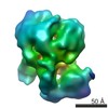
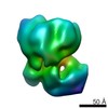
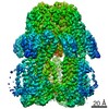
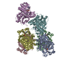
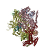
| Title | Electron cryo-microscopy of the HerA-NurA double strand break resection complex | |||||||||
 Map data Map data | Reconstruction of the HerA-NurA double strand break resection complex from Sulfolobus solfataricus | |||||||||
 Sample Sample |
| |||||||||
 Keywords Keywords | Helicase / Nuclease / FtsK/HerA ATPase / DNA double-strand break repair / DNA resection | |||||||||
| Function / homology |  Function and homology information Function and homology informationDNA 5'-3' helicase / exonuclease activity / 3'-5' DNA helicase activity / DNA 3'-5' helicase / endonuclease activity / 5'-3' DNA helicase activity / Hydrolases; Acting on ester bonds / DNA repair / ATP hydrolysis activity / DNA binding ...DNA 5'-3' helicase / exonuclease activity / 3'-5' DNA helicase activity / DNA 3'-5' helicase / endonuclease activity / 5'-3' DNA helicase activity / Hydrolases; Acting on ester bonds / DNA repair / ATP hydrolysis activity / DNA binding / ATP binding / metal ion binding Similarity search - Function | |||||||||
| Biological species |   Sulfolobus solfataricus (archaea) Sulfolobus solfataricus (archaea) | |||||||||
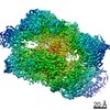
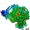
| Method | single particle reconstruction / cryo EM / Resolution: 7.35 Å | |||||||||
 Authors Authors | Byrne RT / Schuller JM / Unverdorben P / Foerster F / Hopfner K-P | |||||||||
 Citation Citation | Journal: FEBS Lett / Year: 2014 Title: Molecular architecture of the HerA-NurA DNA double-strand break resection complex. Authors: Robert Thomas Byrne / Jan Michael Schuller / Pia Unverdorben / Friedrich Förster / Karl-Peter Hopfner /  Abstract: DNA double-strand breaks can be repaired by homologous recombination, during which the DNA ends are long-range resected by helicase-nuclease systems to generate 3' single strand tails. In archaea, ...DNA double-strand breaks can be repaired by homologous recombination, during which the DNA ends are long-range resected by helicase-nuclease systems to generate 3' single strand tails. In archaea, this requires the Mre11-Rad50 complex and the ATP-dependent helicase-nuclease complex HerA-NurA. We report the cryo-EM structure of Sulfolobus solfataricus HerA-NurA at 7.4Å resolution and present the pseudo-atomic model of the complex. HerA forms an ASCE hexamer that tightly interacts with a NurA dimer, with each NurA protomer binding three adjacent HerA HAS domains. Entry to NurA's nuclease active sites requires dsDNA to pass through a 23Å wide channel in the HerA hexamer. The structure suggests that HerA is a dsDNA translocase that feeds DNA into the NurA nuclease sites. | |||||||||
| History |
|
- Structure visualization
| Movie |
Movie viewer |
|---|---|
| Structure viewer | EM map: SurfViewMolmilJmol/JSmol |
| Supplemental images |


- Downloads & links
-EMDB archive
| Map data | emd_2808.map.gz | 2.1 MB | EMDB map data format | |
|---|---|---|---|---|
| Header (meta data) | emd-2808-v30.xmlemd-2808.xml | 10.8 KB 10.8 KB | Display Display | EMDB header |
| Images | EMD-2808.tifemd_2808.tif | 1 MB 1 MB | ||
| Archive directory |  http://ftp.pdbj.org/pub/emdb/structures/EMD-2808ftp://ftp.pdbj.org/pub/emdb/structures/EMD-2808 http://ftp.pdbj.org/pub/emdb/structures/EMD-2808ftp://ftp.pdbj.org/pub/emdb/structures/EMD-2808 | HTTPS FTP |
-Related structure data
| Similar structure data |
|---|
-Links
| EMDB pages | EMDB (EBI/PDBe) / EMDataResource |
|---|
-Map
| File | Download / File: emd_2808.map.gz / Format: CCP4 / Size: 12.6 MB / Type: IMAGE STORED AS FLOATING POINT NUMBER (4 BYTES) | ||||||||||||||||||||||||||||||||||||||||||||||||||||||||||||||||||||
|---|---|---|---|---|---|---|---|---|---|---|---|---|---|---|---|---|---|---|---|---|---|---|---|---|---|---|---|---|---|---|---|---|---|---|---|---|---|---|---|---|---|---|---|---|---|---|---|---|---|---|---|---|---|---|---|---|---|---|---|---|---|---|---|---|---|---|---|---|---|
| Annotation | Reconstruction of the HerA-NurA double strand break resection complex from Sulfolobus solfataricus | ||||||||||||||||||||||||||||||||||||||||||||||||||||||||||||||||||||
| Projections & slices | Image control
Images are generated by Spider. | ||||||||||||||||||||||||||||||||||||||||||||||||||||||||||||||||||||
| Voxel size | X=Y=Z: 1.77 Å | ||||||||||||||||||||||||||||||||||||||||||||||||||||||||||||||||||||
| Density |
| ||||||||||||||||||||||||||||||||||||||||||||||||||||||||||||||||||||
| Symmetry | Space group: 1 | ||||||||||||||||||||||||||||||||||||||||||||||||||||||||||||||||||||
| Details | EMDB XML:
CCP4 map header:
| ||||||||||||||||||||||||||||||||||||||||||||||||||||||||||||||||||||
 Z (Sec.)
 Y (Row.)
 X (Col.)


-Supplemental data
- Sample components
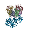
-Entire : HerA from Sulfolobus Solfataricus NurA from Sulfolobus Solfataricus
| Entire | Name: HerA from Sulfolobus Solfataricus NurA from Sulfolobus Solfataricus |
|---|---|
| Components |
|
-Supramolecule #1000: HerA from Sulfolobus Solfataricus NurA from Sulfolobus Solfataricus
| Supramolecule | Name: HerA from Sulfolobus Solfataricus NurA from Sulfolobus Solfataricus type: sample / ID: 1000 / Details: The sample was monodisperse Oligomeric state: One homohexamer of HerA binds to one homodimer of NurA Number unique components: 2 |
|---|---|
| Molecular weight | Experimental: 419 KDa / Theoretical: 416 KDa / Method: SEC-RALS |
-Macromolecule #1: HerA
| Macromolecule | Name: HerA / type: protein_or_peptide / ID: 1 / Name.synonym: SSO2251 / Number of copies: 6 / Oligomeric state: homohexamer / Recombinant expression: Yes |
|---|---|
| Source (natural) | Organism: Sulfolobus solfataricus (archaea) / Strain: P2 |
| Molecular weight | Theoretical: 56 KDa |
| Recombinant expression | Organism:  |
| Sequence | UniProtKB: DNA double-strand break repair helicase HerA |
-Macromolecule #2: NurA
| Macromolecule | Name: NurA / type: protein_or_peptide / ID: 2 / Name.synonym: SSO2248 / Number of copies: 2 / Oligomeric state: dimer / Recombinant expression: Yes |
|---|---|
| Source (natural) | Organism: Sulfolobus solfataricus (archaea) / Strain: P2 |
| Molecular weight | Theoretical: 39 KDa |
| Recombinant expression | Organism: |
| Sequence | UniProtKB: DNA double-strand break repair nuclease NurA |
-Experimental details
-Structure determination
| Method | cryo EM |
|---|---|
 Processing Processing | single particle reconstruction |
| Aggregation state | particle |
-Sample preparation
| Concentration | 0.05 mg/mL |
|---|---|
| Buffer | pH: 8 / Details: 100 mM NaCl, 20 mM Hepes |
| Grid | Details: 2:1 holey carbon grids (Quantifoil Micro Tools, Germany) |
| Vitrification | Cryogen name: ETHANE / Chamber temperature: 93 K / Instrument: HOMEMADE PLUNGER Method: Blot for 2 seconds before plunging, wash twice with water |
- Electron microscopy
| Microscope | FEI TITAN KRIOS |
|---|---|
| Alignment procedure | Legacy - Astigmatism: Objective lens astigmatism was corrected at 100,000 times magnification |
| Specialist optics | Energy filter - Name: GIF QUANTUM / Energy filter - Lower energy threshold: 0.0 eV / Energy filter - Upper energy threshold: 20.0 eV |
| Date | Aug 22, 2014 |
| Image recording | Category: CCD / Film or detector model: GATAN K2 (4k x 4k) / Number real images: 863 / Average electron dose: 25 e/Å2 Details: Every image is the average of 20 frames recorded by the direct electron detector |
| Electron beam | Acceleration voltage: 300 kV / Electron source:  FIELD EMISSION GUN FIELD EMISSION GUN |
| Electron optics | Illumination mode: FLOOD BEAM / Imaging mode: BRIGHT FIELD / Cs: 2 mm / Nominal defocus max: 3.0 µm / Nominal defocus min: 1.5 µm / Nominal magnification: 81000 |
| Sample stage | Specimen holder model: FEI TITAN KRIOS AUTOGRID HOLDER |
| Experimental equipment |  Model: Titan Krios / Image courtesy: FEI Company |
-Image processing
| Details | The particles were semi-manually selected in e2boxer and further processed in RELION |
|---|---|
| CTF correction | Details: On the micrograph level |
| Final reconstruction | Applied symmetry - Point group: C2 (2 fold cyclic) / Algorithm: OTHER / Resolution.type: BY AUTHOR / Resolution: 7.35 Å / Resolution method: OTHER / Software - Name: RELION / Details: see detailed method in paper / Number images used: 100000 |
