| Entry | Database: PDB / ID: 6gx1
|
|---|
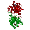
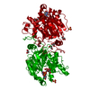
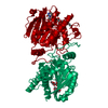
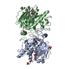
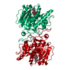
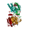
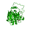
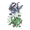
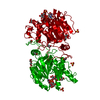
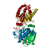
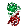
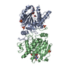
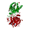
| Title | Blood group synthase AAGlyB in complex with UDP-GalNAc and cryoprotected with PEG 3350 |
|---|
 Components Components | ABO blood group (transferase A, alpha 1-3-N-acetylgalactosaminyltransferase transferase B, alpha 1-3-galactosyltransferase) |
|---|
 Keywords Keywords | TRANSFERASE / blood group synthase / glycosyltransferase / dual specificity / cis-AB mutant |
|---|
| Function / homology |  Function and homology information Function and homology information
fucosylgalactoside 3-alpha-galactosyltransferase / glycoprotein-fucosylgalactoside alpha-N-acetylgalactosaminyltransferase / glycoprotein-fucosylgalactoside alpha-N-acetylgalactosaminyltransferase activity / fucosylgalactoside 3-alpha-galactosyltransferase activity / ABO blood group biosynthesis / hexosyltransferase activity / Golgi cisterna membrane / antigen binding / manganese ion binding / carbohydrate metabolic process ...fucosylgalactoside 3-alpha-galactosyltransferase / glycoprotein-fucosylgalactoside alpha-N-acetylgalactosaminyltransferase / glycoprotein-fucosylgalactoside alpha-N-acetylgalactosaminyltransferase activity / fucosylgalactoside 3-alpha-galactosyltransferase activity / ABO blood group biosynthesis / hexosyltransferase activity / Golgi cisterna membrane / antigen binding / manganese ion binding / carbohydrate metabolic process / vesicle / Golgi membrane / nucleotide binding / Golgi apparatus / extracellular region / membrane / metal ion bindingSimilarity search - Function Glycosyl transferase, family 6 / Glycosyltransferase family 6 / Spore Coat Polysaccharide Biosynthesis Protein SpsA; Chain A / Spore Coat Polysaccharide Biosynthesis Protein SpsA; Chain A / Nucleotide-diphospho-sugar transferases / Alpha-Beta Complex / Alpha BetaSimilarity search - Domain/homology : / DI(HYDROXYETHYL)ETHER / URIDINE-DIPHOSPHATE-N-ACETYLGALACTOSAMINE / URIDINE-5'-DIPHOSPHATE / ABO blood group (transferase A, alpha 1-3-N-acetylgalactosaminyltransferase transferase B, alpha 1-3-galactosyltransferase) / Histo-blood group ABO system transferaseSimilarity search - Component |
|---|
| Biological species |  Homo sapiens (human) Homo sapiens (human) |
|---|
| Method |  X-RAY DIFFRACTION / SYNCHROTRON / FOURIER SYNTHESIS / Resolution: 1.6 Å X-RAY DIFFRACTION / SYNCHROTRON / FOURIER SYNTHESIS / Resolution: 1.6 Å |
|---|
 Authors Authors | Rocha, J. / Royant, A. |
|---|
| Funding support |  France, 1items France, 1items | Organization | Grant number | Country |
|---|
| French National Research Agency | ANR-13-BSV8-0011-02 | France |
|
|---|
 Citation Citation | Journal: To be published
Title: Blood group synthase AAGlyB in complex with UDP-GalNAc and cryoprotected with PEG 3350
Authors: Rocha, J. / Batot, G.O. / Palcic, M.M. / Breton, C. / Royant, A. |
|---|
| History | | Deposition | Jun 26, 2018 | Deposition site: PDBE / Processing site: PDBE |
|---|
| Revision 1.0 | Jul 10, 2019 | Provider: repository / Type: Initial release |
|---|
| Revision 1.1 | Jan 17, 2024 | Group: Data collection / Database references ...Data collection / Database references / Derived calculations / Refinement description
Category: chem_comp_atom / chem_comp_bond ...chem_comp_atom / chem_comp_bond / database_2 / pdbx_initial_refinement_model / struct_conn
Item: _database_2.pdbx_DOI / _database_2.pdbx_database_accession ..._database_2.pdbx_DOI / _database_2.pdbx_database_accession / _struct_conn.pdbx_dist_value / _struct_conn.pdbx_ptnr1_label_alt_id / _struct_conn.pdbx_ptnr2_label_alt_id / _struct_conn.ptnr2_auth_comp_id / _struct_conn.ptnr2_auth_seq_id / _struct_conn.ptnr2_label_asym_id / _struct_conn.ptnr2_label_atom_id / _struct_conn.ptnr2_label_comp_id |
|---|
|
|---|
 Movie
Movie Controller

 Yorodumi
 Open data
 Basic information
 Structure visualization
 Downloads & links
 Other downloads
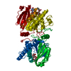
 PDBj
PDBj


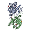
 Assembly
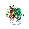


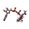


 Mass: 54.938 Da / Num. of mol.: 1 / Source method: obtained synthetically / Formula: Mn
 Mass: 607.354 Da / Num. of mol.: 1 / Source method: obtained synthetically / Formula: C17H27N3O17P2
 Type: RNA linking / Mass: 404.161 Da / Num. of mol.: 1 / Source method: obtained synthetically / Formula: C9H14N2O12P2 / Comment: UDP*YM
 Mass: 96.063 Da / Num. of mol.: 3 / Source method: obtained synthetically / Formula: SO4
 Mass: 106.120 Da / Num. of mol.: 1 / Source method: obtained synthetically / Formula: C4H10O3
 Sample preparation
 Processing