 Movie
Movie Controller


+ Open data

- Basic information
| Entry | Database: PDB / ID: 6gto | ||||||
|---|---|---|---|---|---|---|---|
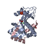
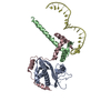
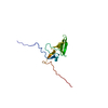
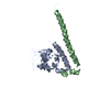
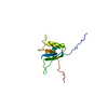
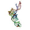
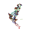
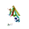
| Title | Structure of the AtaR antitoxin | ||||||
 Components Components | DUF1778 domain-containing protein | ||||||
 Keywords Keywords | TRANSCRIPTION / antitoxin / transcription factor / ribbon-helix-helix / RHH / bacterial repressor | ||||||
| Function / homology | Vibrio phage ICP1, Orf50 / Antitoxin TacA 1-like / toxin sequestering activity / Ribbon-helix-helix / regulation of DNA-templated transcription / DUF1778 domain-containing protein Function and homology information Function and homology information | ||||||
| Biological species |  | ||||||
| Method |  X-RAY DIFFRACTION / SYNCHROTRON / MOLECULAR REPLACEMENT / Resolution: 2.97 Å X-RAY DIFFRACTION / SYNCHROTRON / MOLECULAR REPLACEMENT / Resolution: 2.97 Å | ||||||
 Authors Authors | Garcia-Pino, A. / Jurenas, D. | ||||||
 Citation Citation | Journal: Nat. Chem. Biol. / Year: 2019 Title: Mechanism of regulation and neutralization of the AtaR-AtaT toxin-antitoxin system. Authors: Jurenas, D. / Van Melderen, L. / Garcia-Pino, A. | ||||||
| History |
|
- Structure visualization
| Structure viewer | Molecule: MolmilJmol/JSmol |
|---|
- Downloads & links
-Download
| PDBx/mmCIF format | 6gto.cif.gz | 38 KB | Display | PDBx/mmCIF format |
|---|---|---|---|---|
| PDB format | pdb6gto.ent.gz | 25.4 KB | Display | PDB format |
| PDBx/mmJSON format | 6gto.json.gz | Tree view | PDBx/mmJSON format | |
| Others |  Other downloads Other downloads |
-Validation report
| Arichive directory | https://data.pdbj.org/pub/pdb/validation_reports/gt/6gtoftp://data.pdbj.org/pub/pdb/validation_reports/gt/6gto | HTTPS FTP |
|---|
-Related structure data


-Links
 PDBj
PDBj- Assembly
| Deposited unit | 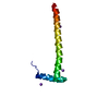
| ||||||||
|---|---|---|---|---|---|---|---|---|---|
| 1 | 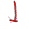
| ||||||||
| Unit cell |
|
-Components
| #1: Protein | Mass: 9925.330 Da / Num. of mol.: 1 Source method: isolated from a genetically manipulated source Source: (gene. exp.) Gene: ybl13, ybl13_1, A8G17_04185, AA102_09360, AC789_1c38270, ACN002_3543, ACN81_27750, ACU90_15595, AM270_07745, ARC77_13665, AU473_04475, B1K96_23180, B1K96_30445, BHS81_20640, BIZ41_06975, BK292_ ...Gene: ybl13, ybl13_1, A8G17_04185, AA102_09360, AC789_1c38270, ACN002_3543, ACN81_27750, ACU90_15595, AM270_07745, ARC77_13665, AU473_04475, B1K96_23180, B1K96_30445, BHS81_20640, BIZ41_06975, BK292_07330, BMT53_16880, BN17_33961, BTQ04_07040, BWP17_00645, COD46_18440, CR538_01655, CVH05_12360, ERS085366_00076, ERS085374_01548, ERS085383_01733, ERS085404_01502, RX35_03224 Production host: References: UniProt: J7QA90 |
|---|---|
| #2: Chemical | ChemComp-NA /   Mass: 22.990 Da / Num. of mol.: 4 / Source method: obtained synthetically / Formula: Na Mass: 22.990 Da / Num. of mol.: 4 / Source method: obtained synthetically / Formula: Na |
-Experimental details
-Experiment
| Experiment | Method: X-RAY DIFFRACTION / Number of used crystals: 1 |
|---|
- Sample preparation
| Crystal | Density Matthews: 3.78 Å3/Da / Density % sol: 67.47 % |
|---|---|
| Crystal grow | Temperature: 298 K / Method: vapor diffusion, sitting drop / pH: 7 / Details: 35%(v/v) 1,4-dioxane |
-Data collection
| Diffraction | Mean temperature: 100 K |
|---|---|
| Diffraction source | Source: SYNCHROTRON / Site: SOLEIL  / Beamline: PROXIMA 1 / Wavelength: 0.9793 Å / Beamline: PROXIMA 1 / Wavelength: 0.9793 Å |
| Detector | Type: DECTRIS PILATUS 6M / Detector: PIXEL / Date: May 18, 2015 |
| Radiation | Protocol: SINGLE WAVELENGTH / Monochromatic (M) / Laue (L): M / Scattering type: x-ray |
| Radiation wavelength | Wavelength: 0.9793 Å / Relative weight: 1 |
| Reflection | Resolution: 2.97→41.86 Å / Num. obs: 3575 / % possible obs: 99.61 % / Redundancy: 5.5 % / Biso Wilson estimate: 117.7 Å2 / Rmerge(I) obs: 0.056 / Net I/σ(I): 9.58 |
| Reflection shell | Resolution: 2.97→3.08 Å / Rmerge(I) obs: 1.482 / CC1/2: 0.538 / % possible all: 99.42 |
- Processing
| Software |
| ||||||||||||||||||||||||||||||||||||||||||||||||||||||||||||||||||||||||||||||||||||||||||||||||||||||||||||||||||
|---|---|---|---|---|---|---|---|---|---|---|---|---|---|---|---|---|---|---|---|---|---|---|---|---|---|---|---|---|---|---|---|---|---|---|---|---|---|---|---|---|---|---|---|---|---|---|---|---|---|---|---|---|---|---|---|---|---|---|---|---|---|---|---|---|---|---|---|---|---|---|---|---|---|---|---|---|---|---|---|---|---|---|---|---|---|---|---|---|---|---|---|---|---|---|---|---|---|---|---|---|---|---|---|---|---|---|---|---|---|---|---|---|---|---|---|
| Refinement | Method to determine structure: MOLECULAR REPLACEMENT / Resolution: 2.97→41.86 Å / Cor.coef. Fo:Fc: 0.93 / Cor.coef. Fo:Fc free: 0.893 / SU R Cruickshank DPI: 0.359 / Cross valid method: THROUGHOUT / σ(F): 0 / SU R Blow DPI: 0.4 / SU Rfree Blow DPI: 0.295 / SU Rfree Cruickshank DPI: 0.283
| ||||||||||||||||||||||||||||||||||||||||||||||||||||||||||||||||||||||||||||||||||||||||||||||||||||||||||||||||||
| Displacement parameters | Biso mean: 134.29 Å2
| ||||||||||||||||||||||||||||||||||||||||||||||||||||||||||||||||||||||||||||||||||||||||||||||||||||||||||||||||||
| Refine analyze | Luzzati coordinate error obs: 0.56 Å | ||||||||||||||||||||||||||||||||||||||||||||||||||||||||||||||||||||||||||||||||||||||||||||||||||||||||||||||||||
| Refinement step | Cycle: 1 / Resolution: 2.97→41.86 Å
| ||||||||||||||||||||||||||||||||||||||||||||||||||||||||||||||||||||||||||||||||||||||||||||||||||||||||||||||||||
| Refine LS restraints |
| ||||||||||||||||||||||||||||||||||||||||||||||||||||||||||||||||||||||||||||||||||||||||||||||||||||||||||||||||||
| LS refinement shell | Resolution: 2.97→3.32 Å / Total num. of bins used: 5
| ||||||||||||||||||||||||||||||||||||||||||||||||||||||||||||||||||||||||||||||||||||||||||||||||||||||||||||||||||
| Refinement TLS params. | Method: refined / Origin x: 27.1228 Å / Origin y: 19.2179 Å / Origin z: 75.0485 Å
| ||||||||||||||||||||||||||||||||||||||||||||||||||||||||||||||||||||||||||||||||||||||||||||||||||||||||||||||||||
| Refinement TLS group | Selection details: { A|* } |
