 Movie
Movie Controller

[English] 日本語
 Yorodumi
- PDB-6gtj: Neutron crystal structure for copper nitrite reductase from Achro... -
+ Open data

- Basic information
| Entry | Database: PDB / ID: 6gtj | ||||||||||||
|---|---|---|---|---|---|---|---|---|---|---|---|---|---|
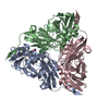
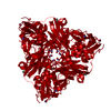
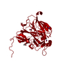
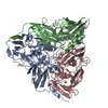
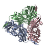
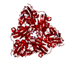
| Title | Neutron crystal structure for copper nitrite reductase from Achromobacter Cycloclastes at 1.8 A resolution | ||||||||||||
 Components Components | Copper-containing nitrite reductase | ||||||||||||
 Keywords Keywords | OXIDOREDUCTASE / Perdueteration / NITRITE REDUCTASE / electron transfer | ||||||||||||
| Function / homology |  Function and homology information Function and homology informationdenitrification pathway / nitrite reductase (NO-forming) / nitrite reductase (NO-forming) activity / nitrate assimilation / periplasmic space / copper ion binding Similarity search - Function | ||||||||||||
| Biological species |  Achromobacter cycloclastes (bacteria) Achromobacter cycloclastes (bacteria) | ||||||||||||
| Method | NEUTRON DIFFRACTION / NUCLEAR REACTOR /  MOLECULAR REPLACEMENT / Resolution: 1.801 Å MOLECULAR REPLACEMENT / Resolution: 1.801 Å | ||||||||||||
 Authors Authors | Antonyuk, S.V. / Blakeley, M.P. / Halsted, T.P. / Eady, R.R. / Hasnain, S.S. | ||||||||||||
| Funding support |  United Kingdom, 3items United Kingdom, 3items
| ||||||||||||
 Citation Citation | Journal: Iucrj / Year: 2019 Title: Catalytically important damage-free structures of a copper nitrite reductase obtained by femtosecond X-ray laser and room-temperature neutron crystallography. Authors: Halsted, T.P. / Yamashita, K. / Gopalasingam, C.C. / Shenoy, R.T. / Hirata, K. / Ago, H. / Ueno, G. / Blakeley, M.P. / Eady, R.R. / Antonyuk, S.V. / Yamamoto, M. / Hasnain, S.S. | ||||||||||||
| History |
|
- Structure visualization
| Structure viewer | Molecule: MolmilJmol/JSmol |
|---|
- Downloads & links
-Download
| PDBx/mmCIF format | 6gtj.cif.gz | 143.6 KB | Display | PDBx/mmCIF format |
|---|---|---|---|---|
| PDB format | pdb6gtj.ent.gz | 113.7 KB | Display | PDB format |
| PDBx/mmJSON format | 6gtj.json.gz | Tree view | PDBx/mmJSON format | |
| Others |  Other downloads Other downloads |
-Validation report
| Arichive directory | https://data.pdbj.org/pub/pdb/validation_reports/gt/6gtjftp://data.pdbj.org/pub/pdb/validation_reports/gt/6gtj | HTTPS FTP |
|---|
-Related structure data
| Related structure data |  6gsqC  6gt0C  6gt2C  6gtiC  6gtkC  6gtlC  6gtnC  2bw4S S: Starting model for refinement C: citing same article ( |
|---|---|
| Similar structure data |


-Links
 PDBj
PDBj

- Assembly
| Deposited unit | 
| |||||||||
|---|---|---|---|---|---|---|---|---|---|---|
| 1 | 
| |||||||||
| Unit cell |
| |||||||||
| Components on special symmetry positions |
|
-Components
| #1: Protein | Mass: 37059.809 Da / Num. of mol.: 1 / Fragment: UNP residues 39-378 Source method: isolated from a genetically manipulated source Source: (gene. exp.) Achromobacter cycloclastes (bacteria) / Gene: nirK / Production host: | ||
|---|---|---|---|
| #2: Chemical |   Mass: 63.546 Da / Num. of mol.: 2 / Source method: obtained synthetically / Formula: Cu Mass: 63.546 Da / Num. of mol.: 2 / Source method: obtained synthetically / Formula: Cu#3: Water | ChemComp-HOH / |  Mass: 18.015 Da / Num. of mol.: 190 / Source method: isolated from a natural source / Formula: H2O Mass: 18.015 Da / Num. of mol.: 190 / Source method: isolated from a natural source / Formula: H2O |
-Experimental details
-Experiment
| Experiment | Method: NEUTRON DIFFRACTION / Number of used crystals: 1 |
|---|
- Sample preparation
| Crystal | Density Matthews: 2 Å3/Da / Density % sol: 35 % / Description: pyramidal shape |
|---|---|
| Crystal grow | Temperature: 297 K / Method: vapor diffusion, hanging drop / pH: 5 Details: 1.2M Ammonium sulphate, Citrate buffer pH 5 all deuterium based |
-Data collection
| Diffraction | Mean temperature: 293 K / Serial crystal experiment: N | |||||||||
|---|---|---|---|---|---|---|---|---|---|---|
| Diffraction source | Source: NUCLEAR REACTOR / Site: ILL  / Beamline: LADI III / Wavelength: 3.05 - 4.00 / Beamline: LADI III / Wavelength: 3.05 - 4.00 | |||||||||
| Detector | Type: LADI III / Detector: DIFFRACTOMETER / Date: Jul 24, 2015 | |||||||||
| Radiation | Protocol: LAUE / Monochromatic (M) / Laue (L): L / Scattering type: neutron | |||||||||
| Radiation wavelength |
| |||||||||
| Reflection | Resolution: 1.8→40 Å / Num. obs: 24728 / % possible obs: 85.5 % / Redundancy: 6.5 % / Rpim(I) all: 0.063 / Net I/σ(I): 7.9 | |||||||||
| Reflection shell | Resolution: 1.8→1.9 Å / Redundancy: 2.9 % / Mean I/σ(I) obs: 3.7 / Num. unique obs: 1858 / Rpim(I) all: 0.13 / % possible all: 69.8 |
- Processing
| Software |
| ||||||||||||||||||||||||
|---|---|---|---|---|---|---|---|---|---|---|---|---|---|---|---|---|---|---|---|---|---|---|---|---|---|
| Refinement | Method to determine structure: MOLECULAR REPLACEMENT Starting model: 2BW4 Resolution: 1.801→32.66 Å / SU ML: 0.25 / Cross valid method: FREE R-VALUE / σ(F): 1.56 / Phase error: 26.04
| ||||||||||||||||||||||||
| Solvent computation | Shrinkage radii: 0.9 Å / VDW probe radii: 1.11 Å | ||||||||||||||||||||||||
| Refine LS restraints |
| ||||||||||||||||||||||||
| LS refinement shell | Resolution: 1.801→1.8825 Å
|
