 Movie
Movie Controller

[English] 日本語
 Yorodumi
- PDB-6gdr: DNA binding with a minimal scaffold: Structure-function analysis ... -
+ Open data

- Basic information
| Entry | Database: PDB / ID: 6gdr | ||||||
|---|---|---|---|---|---|---|---|
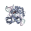
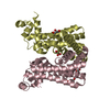
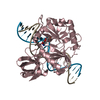
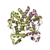
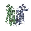
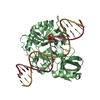
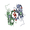
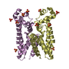
| Title | DNA binding with a minimal scaffold: Structure-function analysis of Lig E DNA ligases | ||||||
 Components Components |
| ||||||
 Keywords Keywords | LIGASE / DNA ligases | ||||||
| Function / homology |  Function and homology information Function and homology informationDNA ligase (ATP) activity / DNA recombination / DNA repair / ATP binding Similarity search - Function | ||||||
| Biological species |  Alteromonas mediterranea (bacteria) Alteromonas mediterranea (bacteria) | ||||||
| Method |  X-RAY DIFFRACTION / SYNCHROTRON / MOLECULAR REPLACEMENT / Resolution: 2.33 Å X-RAY DIFFRACTION / SYNCHROTRON / MOLECULAR REPLACEMENT / Resolution: 2.33 Å | ||||||
 Authors Authors | Williamson, A. / Grigic, M. / Leiros, H.K.S. | ||||||
| Funding support |  Norway, 1items Norway, 1items
| ||||||
 Citation Citation | Journal: Nucleic Acids Res. / Year: 2018 Title: DNA binding with a minimal scaffold: structure-function analysis of Lig E DNA ligases. Authors: Williamson, A. / Grgic, M. / Leiros, H.S. | ||||||
| History |
|
- Structure visualization
| Structure viewer | Molecule: MolmilJmol/JSmol |
|---|
- Downloads & links
-Download
| PDBx/mmCIF format | 6gdr.cif.gz | 149.3 KB | Display | PDBx/mmCIF format |
|---|---|---|---|---|
| PDB format | pdb6gdr.ent.gz | 114.3 KB | Display | PDB format |
| PDBx/mmJSON format | 6gdr.json.gz | Tree view | PDBx/mmJSON format | |
| Others |  Other downloads Other downloads |
-Validation report
| Arichive directory | https://data.pdbj.org/pub/pdb/validation_reports/gd/6gdrftp://data.pdbj.org/pub/pdb/validation_reports/gd/6gdr | HTTPS FTP |
|---|
-Related structure data
| Similar structure data |
|---|

-Links
 PDBj
PDBj


- Assembly
| Deposited unit | 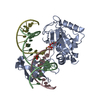
| ||||||||
|---|---|---|---|---|---|---|---|---|---|
| 1 |
| ||||||||
| Unit cell |
|
-Components
-DNA chain , 3 types, 3 molecules BCD
| #1: DNA chain | Mass: 6494.194 Da / Num. of mol.: 1 Source method: isolated from a genetically manipulated source Source: (gene. exp.) Alteromonas mediterranea (bacteria) / Production host: |
|---|---|
| #2: DNA chain | Mass: 3004.981 Da / Num. of mol.: 1 Source method: isolated from a genetically manipulated source Source: (gene. exp.) Alteromonas mediterranea (bacteria) / Production host: |
| #3: DNA chain | Mass: 3342.212 Da / Num. of mol.: 1 Source method: isolated from a genetically manipulated source Source: (gene. exp.) Alteromonas mediterranea (bacteria) / Production host: |
-Protein , 1 types, 1 molecules A
| #4: Protein | Mass: 33017.656 Da / Num. of mol.: 1 Source method: isolated from a genetically manipulated source Source: (gene. exp.) Alteromonas mediterranea (bacteria) / Gene: BM525_03130 / Production host: |
|---|
-Non-polymers , 3 types, 72 molecules 


| #5: Chemical |  Mass: 94.971 Da / Num. of mol.: 2 / Source method: obtained synthetically / Formula: PO4 Mass: 94.971 Da / Num. of mol.: 2 / Source method: obtained synthetically / Formula: PO4#6: Chemical | ChemComp-AMP / |  Mass: 347.221 Da / Num. of mol.: 1 / Source method: obtained synthetically / Formula: C10H14N5O7P / Comment: AMP*YM Mass: 347.221 Da / Num. of mol.: 1 / Source method: obtained synthetically / Formula: C10H14N5O7P / Comment: AMP*YM#7: Water | ChemComp-HOH / | Mass: 18.015 Da / Num. of mol.: 69 / Source method: isolated from a natural source / Formula: H2O |
|---|
-Experimental details
-Experiment
| Experiment | Method: X-RAY DIFFRACTION / Number of used crystals: 1 |
|---|
- Sample preparation
| Crystal | Density Matthews: 2.94 Å3/Da / Density % sol: 58.23 % |
|---|---|
| Crystal grow | Temperature: 277 K / Method: vapor diffusion, hanging drop Details: 24% PEG 4K, 100 mM Bis-Tris pH 5.5, 12% ethyleneglycol |
-Data collection
| Diffraction | Mean temperature: 100 K |
|---|---|
| Diffraction source | Source: SYNCHROTRON / Site: BESSY  / Beamline: 14.1 / Wavelength: 0.9184 Å / Beamline: 14.1 / Wavelength: 0.9184 Å |
| Detector | Type: DECTRIS PILATUS3 S 6M / Detector: PIXEL / Date: Nov 25, 2016 |
| Radiation | Protocol: SINGLE WAVELENGTH / Monochromatic (M) / Laue (L): M / Scattering type: x-ray |
| Radiation wavelength | Wavelength: 0.9184 Å / Relative weight: 1 |
| Reflection | Resolution: 2.33→47.92 Å / Num. obs: 22739 / % possible obs: 98.8 % / Observed criterion σ(F): 0 / Redundancy: 6.9 % / Biso Wilson estimate: 39.4 Å2 / CC1/2: 0.999 / Rmerge(I) obs: 0.075 / Rpim(I) all: 0.046 / Rrim(I) all: 0.088 / Net I/σ(I): 14.6 |
| Reflection shell | Resolution: 2.33→2.41 Å / Redundancy: 6.9 % / Rmerge(I) obs: 1.387 / Mean I/σ(I) obs: 1.2 / Num. unique obs: 2184 / CC1/2: 0.493 / Rpim(I) all: 0.0858 / Rrim(I) all: 1.635 / % possible all: 96.1 |
- Processing
| Software |
| |||||||||||||||||||||||||||||||||||||||||||||||||||||||||||||||
|---|---|---|---|---|---|---|---|---|---|---|---|---|---|---|---|---|---|---|---|---|---|---|---|---|---|---|---|---|---|---|---|---|---|---|---|---|---|---|---|---|---|---|---|---|---|---|---|---|---|---|---|---|---|---|---|---|---|---|---|---|---|---|---|---|
| Refinement | Method to determine structure: MOLECULAR REPLACEMENT Starting model: Homology model Resolution: 2.33→24.84 Å / SU ML: 0.39 / Cross valid method: FREE R-VALUE / σ(F): 1.36 / Phase error: 31.17
| |||||||||||||||||||||||||||||||||||||||||||||||||||||||||||||||
| Solvent computation | Shrinkage radii: 0.9 Å / VDW probe radii: 1.11 Å | |||||||||||||||||||||||||||||||||||||||||||||||||||||||||||||||
| Refinement step | Cycle: LAST / Resolution: 2.33→24.84 Å
| |||||||||||||||||||||||||||||||||||||||||||||||||||||||||||||||
| Refine LS restraints |
| |||||||||||||||||||||||||||||||||||||||||||||||||||||||||||||||
| LS refinement shell |
|
