Method to determine structure: AB INITIO PHASING / Resolution: 1.3→46.32 Å / Cor.coef. Fo:Fc: 0.982 / Cor.coef. Fo:Fc free: 0.977 / SU B: 1.349 / SU ML: 0.025 / Cross valid method: THROUGHOUT / ESU R: 0.047 / ESU R Free: 0.045 Details: HYDROGENS HAVE BEEN ADDED IN THE RIDING POSITIONS U VALUES : REFINED INDIVIDUALLY
Rfactor
Num. reflection
% reflection
Selection details
Rfree
0.15325
4375
4.9 %
RANDOM
Rwork
0.12535
-
-
-
obs
0.12673
84383
85.44 %
-
Solvent computation
Ion probe radii: 0.9 Å / Shrinkage radii: 0.9 Å / VDW probe radii: 1.3 Å
Displacement parameters
Biso mean: 23.273 Å2
Baniso -1
Baniso -2
Baniso -3
1-
-0.28 Å2
0 Å2
-0.14 Å2
2-
-
0.11 Å2
0 Å2
3-
-
-
0.19 Å2
Refinement step
Cycle: LAST / Resolution: 1.3→46.32 Å
Protein
Nucleic acid
Ligand
Solvent
Total
Num. atoms
2830
0
32
429
3291
Refine LS restraints
Refine-ID
Type
Dev ideal
Dev ideal target
Number
X-RAY DIFFRACTION
r_bond_refined_d
0.013
0.019
3323
X-RAY DIFFRACTION
r_bond_other_d
0.001
0.02
3167
X-RAY DIFFRACTION
r_angle_refined_deg
1.4
1.971
4538
X-RAY DIFFRACTION
r_angle_other_deg
0.796
3
7376
X-RAY DIFFRACTION
r_dihedral_angle_1_deg
4.163
5
473
X-RAY DIFFRACTION
r_dihedral_angle_2_deg
30.287
23.642
173
X-RAY DIFFRACTION
r_dihedral_angle_3_deg
13.433
15
634
X-RAY DIFFRACTION
r_dihedral_angle_4_deg
15.866
15
47
X-RAY DIFFRACTION
r_chiral_restr
0.088
0.2
494
X-RAY DIFFRACTION
r_gen_planes_refined
0.008
0.02
3931
X-RAY DIFFRACTION
r_gen_planes_other
0.001
0.02
688
X-RAY DIFFRACTION
r_mcbond_it
1.642
2.057
1662
X-RAY DIFFRACTION
r_mcbond_other
1.624
2.051
1659
X-RAY DIFFRACTION
r_mcangle_it
2.129
3.073
2104
X-RAY DIFFRACTION
r_mcangle_other
2.129
3.076
2105
X-RAY DIFFRACTION
r_scbond_it
1.984
2.423
1661
X-RAY DIFFRACTION
r_scbond_other
1.984
2.424
1662
X-RAY DIFFRACTION
r_scangle_other
2.608
3.512
2394
X-RAY DIFFRACTION
r_long_range_B_refined
3.359
26.02
4169
X-RAY DIFFRACTION
r_long_range_B_other
3.285
25.87
4143
X-RAY DIFFRACTION
r_rigid_bond_restr
2.114
3
6489
X-RAY DIFFRACTION
r_sphericity_free
24.243
5
259
X-RAY DIFFRACTION
r_sphericity_bonded
11.515
5
6591
LS refinement shell
Resolution: 1.3→1.334 Å / Total num. of bins used: 20
Rfactor
Num. reflection
% reflection
Rfree
0.256
349
-
Rwork
0.203
6768
-
obs
-
-
92.46 %
+
About Yorodumi
-
News
-
Feb 9, 2022. New format data for meta-information of EMDB entries
New format data for meta-information of EMDB entries
Version 3 of the EMDB header file is now the official format.
The previous official version 1.9 will be removed from the archive.
In the structure databanks used in Yorodumi, some data are registered as the other names, "COVID-19 virus" and "2019-nCoV". Here are the details of the virus and the list of structure data.
Jan 31, 2019. EMDB accession codes are about to change! (news from PDBe EMDB page)
EMDB accession codes are about to change! (news from PDBe EMDB page)
The allocation of 4 digits for EMDB accession codes will soon come to an end. Whilst these codes will remain in use, new EMDB accession codes will include an additional digit and will expand incrementally as the available range of codes is exhausted. The current 4-digit format prefixed with “EMD-” (i.e. EMD-XXXX) will advance to a 5-digit format (i.e. EMD-XXXXX), and so on. It is currently estimated that the 4-digit codes will be depleted around Spring 2019, at which point the 5-digit format will come into force.
The EM Navigator/Yorodumi systems omit the EMD- prefix.
Related info.:Q: What is EMD? / ID/Accession-code notation in Yorodumi/EM Navigator
Yorodumi is a browser for structure data from EMDB, PDB, SASBDB, etc.
This page is also the successor to EM Navigator detail page, and also detail information page/front-end page for Omokage search.
The word "yorodu" (or yorozu) is an old Japanese word meaning "ten thousand". "mi" (miru) is to see.
Related info.:EMDB / PDB / SASBDB / Comparison of 3 databanks / Yorodumi Search / Aug 31, 2016. New EM Navigator & Yorodumi / Yorodumi Papers / Jmol/JSmol / Function and homology information / Changes in new EM Navigator and Yorodumi
 Movie
Movie Controller

 Yorodumi
 Open data
 Basic information
 Components
 Keywords
 Function and homology information
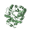
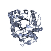
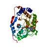
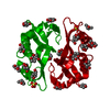
 Pseudomonas aeruginosa (bacteria)
 X-RAY DIFFRACTION /
  Authors
 Spain, 2items
  Citation
 Structure visualization
 Downloads & links
 Other downloads


 PDBj
PDBj


 Assembly

 Mass: 62.005 Da / Num. of mol.: 2 / Source method: obtained synthetically / Formula: NO3 / Feature type: SUBJECT OF INVESTIGATION
 Mass: 22.990 Da / Num. of mol.: 4 / Source method: obtained synthetically / Formula: Na
 Mass: 59.044 Da / Num. of mol.: 5 / Source method: obtained synthetically / Formula: C2H3O2
 Mass: 18.015 Da / Num. of mol.: 429 / Source method: isolated from a natural source / Formula: H2O
 Sample preparation
 / Beamline: ID23-1 / Wavelength: 0.9724 Å
 Processing