 Movie
Movie Controller

[English] 日本語
 Yorodumi
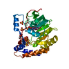
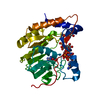
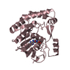
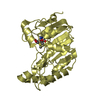
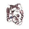
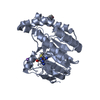
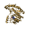
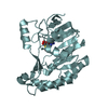
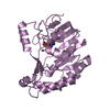
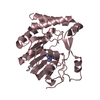
- PDB-3evg: Crystal structure of Dengue-2 virus methyltransferase complexed w... -
+ Open data

- Basic information
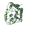
| Entry | Database: PDB / ID: 3evg | ||||||
|---|---|---|---|---|---|---|---|
| Title | Crystal structure of Dengue-2 virus methyltransferase complexed with S-adenosyl-L-homocysteine | ||||||
 Components Components | RNA-directed RNA polymerase NS5 | ||||||
 Keywords Keywords | TRANSFERASE / Dengue virus / Flavivirus / NS5 Methyltransferase / RNA cap binding / ATP-binding / Capsid protein / Cleavage on pair of basic residues / Endoplasmic reticulum / Envelope protein / Glycoprotein / Helicase / Hydrolase / Membrane / Metal-binding / Multifunctional enzyme / Nucleotide-binding / Nucleotidyltransferase / Nucleus / Phosphoprotein / Protease / Ribonucleoprotein / RNA replication / RNA-binding / RNA-directed RNA polymerase / Secreted / Serine protease / Transcription / Transcription regulation / Transmembrane / Viral nucleoprotein / Virion | ||||||
| Function / homology |  Function and homology information Function and homology informationhost cell nucleolus / flavivirin / symbiont-mediated suppression of host JAK-STAT cascade via inhibition of host TYK2 activity / host cell mitochondrion / symbiont-mediated suppression of host JAK-STAT cascade via inhibition of STAT2 activity / symbiont-mediated suppression of host cytoplasmic pattern recognition receptor signaling pathway via inhibition of MAVS activity / viral capsid / nucleoside-triphosphate phosphatase / double-stranded RNA binding / channel activity ...host cell nucleolus / flavivirin / symbiont-mediated suppression of host JAK-STAT cascade via inhibition of host TYK2 activity / host cell mitochondrion / symbiont-mediated suppression of host JAK-STAT cascade via inhibition of STAT2 activity / symbiont-mediated suppression of host cytoplasmic pattern recognition receptor signaling pathway via inhibition of MAVS activity / viral capsid / nucleoside-triphosphate phosphatase / double-stranded RNA binding / channel activity / monoatomic ion transmembrane transport / clathrin-dependent endocytosis of virus by host cell / mRNA (guanine-N7)-methyltransferase / methyltransferase cap1 / methyltransferase cap1 activity / mRNA 5'-cap (guanine-N7-)-methyltransferase activity / RNA helicase activity / protein dimerization activity / host cell perinuclear region of cytoplasm / host cell endoplasmic reticulum membrane / RNA helicase / symbiont-mediated suppression of host type I interferon-mediated signaling pathway / serine-type endopeptidase activity / symbiont-mediated activation of host autophagy / RNA-directed RNA polymerase / viral RNA genome replication / RNA-directed RNA polymerase activity / fusion of virus membrane with host endosome membrane / viral envelope / lipid binding / virion attachment to host cell / DNA-templated transcription / virion membrane / structural molecule activity / ATP hydrolysis activity / proteolysis / extracellular region / ATP binding / metal ion binding Similarity search - Function | ||||||
| Biological species |  Dengue virus 2 16681-PDK53 Dengue virus 2 16681-PDK53 | ||||||
| Method |  X-RAY DIFFRACTION / MOLECULAR REPLACEMENT / Resolution: 2.2 Å X-RAY DIFFRACTION / MOLECULAR REPLACEMENT / Resolution: 2.2 Å | ||||||
 Authors Authors | Thompson, A.A. / Geiss, B.J. / Peersen, O.B. | ||||||
 Citation Citation | Journal: J.Mol.Biol. / Year: 2009 Title: Analysis of flavivirus NS5 methyltransferase cap binding. Authors: Geiss, B.J. / Thompson, A.A. / Andrews, A.J. / Sons, R.L. / Gari, H.H. / Keenan, S.M. / Peersen, O.B. | ||||||
| History |
|
- Structure visualization
| Structure viewer | Molecule: MolmilJmol/JSmol |
|---|
- Downloads & links
-Download
| PDBx/mmCIF format | 3evg.cif.gz | 66.9 KB | Display | PDBx/mmCIF format |
|---|---|---|---|---|
| PDB format | pdb3evg.ent.gz | 48.5 KB | Display | PDB format |
| PDBx/mmJSON format | 3evg.json.gz | Tree view | PDBx/mmJSON format | |
| Others |  Other downloads Other downloads |
-Validation report
| Arichive directory | https://data.pdbj.org/pub/pdb/validation_reports/ev/3evgftp://data.pdbj.org/pub/pdb/validation_reports/ev/3evg | HTTPS FTP |
|---|
-Related structure data
| Related structure data | 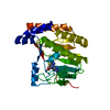 3evaC 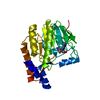 3evbC  3evcC  3evdC  3eveC  3evfC 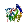 1l9kS C: citing same article ( S: Starting model for refinement |
|---|---|
| Similar structure data |


-Links
 PDBj
PDBj


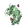
- Assembly
| Deposited unit | 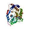
| ||||||||
|---|---|---|---|---|---|---|---|---|---|
| 1 |
| ||||||||
| Unit cell |
|
-Components
| #1: Protein | Mass: 30836.152 Da / Num. of mol.: 1 Fragment: N-terminal domain 1-267 of RNA-directed RNA polymerase NS5: UNP residues 2493-2757 Source method: isolated from a genetically manipulated source Source: (gene. exp.) Dengue virus 2 16681-PDK53 / Strain: 16681 / Plasmid: pKKT7E / Production host:  References: UniProt: P29991, mRNA (guanine-N7)-methyltransferase, RNA-directed RNA polymerase | ||||
|---|---|---|---|---|---|
| #2: Chemical | ChemComp-SO4 /   Mass: 96.063 Da / Num. of mol.: 5 / Source method: obtained synthetically / Formula: SO4 Mass: 96.063 Da / Num. of mol.: 5 / Source method: obtained synthetically / Formula: SO4#3: Chemical | ChemComp-SAH / |   Type: L-peptide linking / Mass: 384.411 Da / Num. of mol.: 1 / Source method: obtained synthetically / Formula: C14H20N6O5S Type: L-peptide linking / Mass: 384.411 Da / Num. of mol.: 1 / Source method: obtained synthetically / Formula: C14H20N6O5S#4: Water | ChemComp-HOH / |  Mass: 18.015 Da / Num. of mol.: 59 / Source method: isolated from a natural source / Formula: H2O Mass: 18.015 Da / Num. of mol.: 59 / Source method: isolated from a natural source / Formula: H2O |
-Experimental details
-Experiment
| Experiment | Method: X-RAY DIFFRACTION / Number of used crystals: 1 |
|---|
- Sample preparation
| Crystal | Density Matthews: 3.31 Å3/Da / Density % sol: 62.8 % |
|---|---|
| Crystal grow | Temperature: 289 K / Method: vapor diffusion, hanging drop / pH: 5 Details: 0.2 M Ammonium sulfate, 0.65 M Lithium sulfate, 0.1 M Sodium citrate pH 5.0, VAPOR DIFFUSION, HANGING DROP, temperature 289K |
-Data collection
| Diffraction | Mean temperature: 100 K |
|---|---|
| Diffraction source | Source: ROTATING ANODE / Type: RIGAKU / Wavelength: 1.54178 Å |
| Detector | Type: RIGAKU RAXIS IV++ / Detector: IMAGE PLATE / Date: May 9, 2006 |
| Radiation | Protocol: SINGLE WAVELENGTH / Monochromatic (M) / Laue (L): M / Scattering type: x-ray |
| Radiation wavelength | Wavelength: 1.54178 Å / Relative weight: 1 |
| Reflection | Resolution: 2.2→28 Å / Num. obs: 39142 / % possible obs: 97.8 % / Redundancy: 6.69 % / Biso Wilson estimate: 18.4 Å2 / Rsym value: 0.099 / Net I/σ(I): 12.7 |
| Reflection shell | Resolution: 2.2→2.28 Å / Redundancy: 6.35 % / Mean I/σ(I) obs: 3.6 / Rsym value: 0.439 / % possible all: 95.1 |
- Processing
| Software |
| |||||||||||||||||||||||||
|---|---|---|---|---|---|---|---|---|---|---|---|---|---|---|---|---|---|---|---|---|---|---|---|---|---|---|
| Refinement | Method to determine structure: MOLECULAR REPLACEMENT Starting model: PDB entry 1L9K Resolution: 2.2→27 Å / Cross valid method: THROUGHOUT / σ(F): 0 / Stereochemistry target values: Engh & Huber Details: The Friedel pairs were used in phasing and refinement
| |||||||||||||||||||||||||
| Displacement parameters | Biso mean: 45.7 Å2
| |||||||||||||||||||||||||
| Refine analyze |
| |||||||||||||||||||||||||
| Refinement step | Cycle: LAST / Resolution: 2.2→27 Å
| |||||||||||||||||||||||||
| Refine LS restraints |
|
