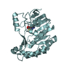
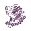
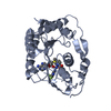
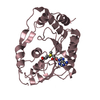
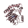
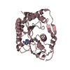
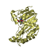
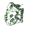
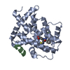
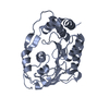
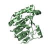
Entry Database : PDB / ID : 4r8rTitle Dengue virus serotype 3 methyltransferase without a bound S-adenosyl methionine nonstructural protein NS5 Keywords / / Function / homology Function Domain/homology Component
/ / / / / / / / / / / / / / / / / / / / / / / / / / / / / / / / / / / / / / / / / / / / / / / / / / / / / / / / / / / / / / / / / / / / / / / / / / / / / / / / / / / / / / / / / / / / / / / / / / / / / / / / / / / / / / / / / / Biological species Method / / / Resolution : 1.46 Å Authors Noble, C.G. Journal : Antiviral Res. / Year : 2014Title : Crystal structure of dengue virus methyltransferase without S-adenosyl-L-methionineAuthors : Noble, C.G. / Li, S.H. / Dong, H. / Chew, S.H. / Shi, P.Y. History Deposition Sep 2, 2014 Deposition site / Processing site Revision 1.0 Oct 15, 2014 Provider / Type Revision 1.1 Nov 8, 2023 Group / Database references / Refinement descriptionCategory chem_comp_atom / chem_comp_bond ... chem_comp_atom / chem_comp_bond / database_2 / pdbx_initial_refinement_model / struct_ref_seq_dif Item / _database_2.pdbx_database_accession / _struct_ref_seq_dif.details
Show all Show less
 Movie
Movie Controller

 Yorodumi
 Open data
 Basic information
 Components
 Keywords
 Function and homology information
 Dengue virus 3
 X-RAY DIFFRACTION /
  Authors
 Citation
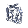
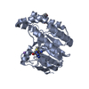
 Structure visualization
 Downloads & links
 Other downloads


 PDBj
PDBj

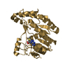
 Assembly
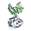

 Mass: 18.015 Da / Num. of mol.: 798 / Source method: isolated from a natural source / Formula: H2O
 Sample preparation
 / Beamline: X10SA / Wavelength: 1 Å
 Processing