 Movie
Movie Controller

[English] 日本語
 Yorodumi
- PDB-6gca: Crystal structure of glutathione transferase Xi 3 from Trametes v... -
+ Open data

- Basic information
| Entry | Database: PDB / ID: 6gca | ||||||
|---|---|---|---|---|---|---|---|
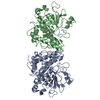
| Title | Crystal structure of glutathione transferase Xi 3 from Trametes versicolor | ||||||
 Components Components | Glutathione transferase Xi 3 from Trametes versicolor | ||||||
 Keywords Keywords | TRANSFERASE / glutathione transferase | ||||||
| Function / homology |  Function and homology information Function and homology information | ||||||
| Biological species |  Trametes versicolor (turkey-tail fungus) Trametes versicolor (turkey-tail fungus) | ||||||
| Method |  X-RAY DIFFRACTION / SYNCHROTRON / MOLECULAR REPLACEMENT / Resolution: 2.282 Å X-RAY DIFFRACTION / SYNCHROTRON / MOLECULAR REPLACEMENT / Resolution: 2.282 Å | ||||||
 Authors Authors | Schwartz, M. / Favier, F. / Didierjean, C. | ||||||
| Funding support |  France, 1items France, 1items
| ||||||
 Citation Citation | Journal: FEBS Lett. / Year: 2018 Title: Trametes versicolor glutathione transferase Xi 3, a dual Cys-GST with catalytic specificities of both Xi and Omega classes. Authors: Schwartz, M. / Perrot, T. / Deroy, A. / Roret, T. / Morel-Rouhier, M. / Mulliert, G. / Gelhaye, E. / Favier, F. / Didierjean, C. | ||||||
| History |
|
- Structure visualization
| Structure viewer | Molecule: MolmilJmol/JSmol |
|---|
- Downloads & links
-Download
| PDBx/mmCIF format | 6gca.cif.gz | 234.6 KB | Display | PDBx/mmCIF format |
|---|---|---|---|---|
| PDB format | pdb6gca.ent.gz | 191.5 KB | Display | PDB format |
| PDBx/mmJSON format | 6gca.json.gz | Tree view | PDBx/mmJSON format | |
| Others |  Other downloads Other downloads |
-Validation report
| Arichive directory | https://data.pdbj.org/pub/pdb/validation_reports/gc/6gcaftp://data.pdbj.org/pub/pdb/validation_reports/gc/6gca | HTTPS FTP |
|---|
-Related structure data
| Related structure data |  6gc9C 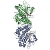 6gcbC 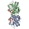 6gccC 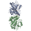 6htaC  3ppuS S: Starting model for refinement C: citing same article ( |
|---|---|
| Similar structure data |


-Links
 PDBj
PDBj

- Assembly
| Deposited unit | 
| ||||||||
|---|---|---|---|---|---|---|---|---|---|
| 1 |
| ||||||||
| Unit cell |
|
-Components
| #1: Protein | Mass: 37163.148 Da / Num. of mol.: 2 Source method: isolated from a genetically manipulated source Source: (gene. exp.) Trametes versicolor (turkey-tail fungus)Production host:  References: UniProt: A0A3F2YM27*PLUS, glutathione transferase #2: Water | ChemComp-HOH / |  Mass: 18.015 Da / Num. of mol.: 355 / Source method: isolated from a natural source / Formula: H2O Mass: 18.015 Da / Num. of mol.: 355 / Source method: isolated from a natural source / Formula: H2O |
|---|
-Experimental details
-Experiment
| Experiment | Method: X-RAY DIFFRACTION / Number of used crystals: 1 |
|---|
- Sample preparation
| Crystal | Density Matthews: 2.55 Å3/Da / Density % sol: 51.83 % |
|---|---|
| Crystal grow | Temperature: 277 K / Method: microbatch / pH: 6.5 Details: 20% (w/v) polyethylene glycol 8,000, 0.1M pH 6.5 sodium cacodylate buffer and 0.2M magnesium acetate |
-Data collection
| Diffraction | Mean temperature: 100 K |
|---|---|
| Diffraction source | Source: SYNCHROTRON / Site: ESRF / Beamline: BM30A / Wavelength: 0.97974 Å |
| Detector | Type: ADSC QUANTUM 315r / Detector: CCD / Date: Nov 15, 2014 |
| Radiation | Protocol: SINGLE WAVELENGTH / Monochromatic (M) / Laue (L): M / Scattering type: x-ray |
| Radiation wavelength | Wavelength: 0.97974 Å / Relative weight: 1 |
| Reflection | Resolution: 2.28→48.68 Å / Num. obs: 33851 / % possible obs: 98.1 % / Redundancy: 3.7 % / Rmerge(I) obs: 0.1 / Net I/σ(I): 14 |
| Reflection shell | Resolution: 2.28→2.34 Å / Rmerge(I) obs: 0.69 / Num. unique all: 2465 |
- Processing
| Software |
| |||||||||||||||||||||||||||||||||||||||||||||||||||||||||||||||||||||||||||||||||||||||||||
|---|---|---|---|---|---|---|---|---|---|---|---|---|---|---|---|---|---|---|---|---|---|---|---|---|---|---|---|---|---|---|---|---|---|---|---|---|---|---|---|---|---|---|---|---|---|---|---|---|---|---|---|---|---|---|---|---|---|---|---|---|---|---|---|---|---|---|---|---|---|---|---|---|---|---|---|---|---|---|---|---|---|---|---|---|---|---|---|---|---|---|---|---|
| Refinement | Method to determine structure: MOLECULAR REPLACEMENT Starting model: 3PPU Resolution: 2.282→36.433 Å / SU ML: 0.29 / Cross valid method: FREE R-VALUE / σ(F): 1.34 / Phase error: 22.79 / Stereochemistry target values: ML
| |||||||||||||||||||||||||||||||||||||||||||||||||||||||||||||||||||||||||||||||||||||||||||
| Solvent computation | Shrinkage radii: 0.9 Å / VDW probe radii: 1.11 Å / Solvent model: FLAT BULK SOLVENT MODEL | |||||||||||||||||||||||||||||||||||||||||||||||||||||||||||||||||||||||||||||||||||||||||||
| Refinement step | Cycle: LAST / Resolution: 2.282→36.433 Å
| |||||||||||||||||||||||||||||||||||||||||||||||||||||||||||||||||||||||||||||||||||||||||||
| Refine LS restraints |
| |||||||||||||||||||||||||||||||||||||||||||||||||||||||||||||||||||||||||||||||||||||||||||
| LS refinement shell |
|
