 Movie
Movie Controller

[English] 日本語
 Yorodumi
- PDB-6gav: Extremely 'open' clamp structure of DNA gyrase: role of the Coryn... -
+ Open data

- Basic information
| Entry | Database: PDB / ID: 6gav | ||||||
|---|---|---|---|---|---|---|---|
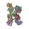
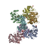
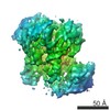
| Title | Extremely 'open' clamp structure of DNA gyrase: role of the Corynebacteriales GyrB specific insert | ||||||
 Components Components | DNA gyrase subunit B,DNA gyrase subunit A | ||||||
 Keywords Keywords | DNA BINDING PROTEIN | ||||||
| Function / homology |  Function and homology information Function and homology informationDNA topoisomerase type II (double strand cut, ATP-hydrolyzing) complex / DNA negative supercoiling activity / DNA topoisomerase type II (double strand cut, ATP-hydrolyzing) activity / DNA topoisomerase (ATP-hydrolysing) / DNA topological change / peptidoglycan-based cell wall / DNA-templated DNA replication / chromosome / response to antibiotic / magnesium ion binding ...DNA topoisomerase type II (double strand cut, ATP-hydrolyzing) complex / DNA negative supercoiling activity / DNA topoisomerase type II (double strand cut, ATP-hydrolyzing) activity / DNA topoisomerase (ATP-hydrolysing) / DNA topological change / peptidoglycan-based cell wall / DNA-templated DNA replication / chromosome / response to antibiotic / magnesium ion binding / ATP hydrolysis activity / DNA binding / ATP binding / metal ion binding / plasma membrane / cytoplasm Similarity search - Function | ||||||
| Biological species |   Mycobacterium tuberculosis (bacteria) Mycobacterium tuberculosis (bacteria) | ||||||
| Method |  X-RAY DIFFRACTION / SYNCHROTRON / MOLECULAR REPLACEMENT / Resolution: 2.6 Å X-RAY DIFFRACTION / SYNCHROTRON / MOLECULAR REPLACEMENT / Resolution: 2.6 Å | ||||||
 Authors Authors | Petrella, S. / Capton, E. / Alzari, P.M. / Aubry, A. / MAyer, C. | ||||||
 Citation Citation | Journal: Structure / Year: 2019 Title: Overall Structures of Mycobacterium tuberculosis DNA Gyrase Reveal the Role of a Corynebacteriales GyrB-Specific Insert in ATPase Activity. Authors: Petrella, S. / Capton, E. / Raynal, B. / Giffard, C. / Thureau, A. / Bonnete, F. / Alzari, P.M. / Aubry, A. / Mayer, C. | ||||||
| History |
|
- Structure visualization
| Structure viewer | Molecule: MolmilJmol/JSmol |
|---|
- Downloads & links
-Download
| PDBx/mmCIF format | 6gav.cif.gz | 880.2 KB | Display | PDBx/mmCIF format |
|---|---|---|---|---|
| PDB format | pdb6gav.ent.gz | 733 KB | Display | PDB format |
| PDBx/mmJSON format | 6gav.json.gz | Tree view | PDBx/mmJSON format | |
| Others |  Other downloads Other downloads |
-Validation report
| Arichive directory | https://data.pdbj.org/pub/pdb/validation_reports/ga/6gavftp://data.pdbj.org/pub/pdb/validation_reports/ga/6gav | HTTPS FTP |
|---|
-Related structure data
| Related structure data |  6gauC  3ifzS 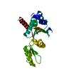 3ig0S  3zkdS S: Starting model for refinement C: citing same article ( |
|---|---|
| Similar structure data |


-Links
 PDBj
PDBj


- Assembly
| Deposited unit | 
| ||||||||
|---|---|---|---|---|---|---|---|---|---|
| 1 |
| ||||||||
| Unit cell |
|
-Components
| #1: Protein | Mass: 130449.359 Da / Num. of mol.: 2 Source method: isolated from a genetically manipulated source Source: (gene. exp.) Mycobacterium tuberculosis (bacteria) / Gene: gyrB, gyrA, Rv0006, MTCY10H4.04 / Production host: #2: Chemical | ChemComp-MES /   Mass: 195.237 Da / Num. of mol.: 5 / Source method: obtained synthetically / Formula: C6H13NO4S / Comment: pH buffer*YM Mass: 195.237 Da / Num. of mol.: 5 / Source method: obtained synthetically / Formula: C6H13NO4S / Comment: pH buffer*YM#3: Water | ChemComp-HOH / |  Mass: 18.015 Da / Num. of mol.: 47 / Source method: isolated from a natural source / Formula: H2O Mass: 18.015 Da / Num. of mol.: 47 / Source method: isolated from a natural source / Formula: H2O |
|---|
-Experimental details
-Experiment
| Experiment | Method: X-RAY DIFFRACTION / Number of used crystals: 1 |
|---|
- Sample preparation
| Crystal | Density Matthews: 3.02 Å3/Da / Density % sol: 59.25 % |
|---|---|
| Crystal grow | Temperature: 290 K / Method: vapor diffusion, sitting drop Details: 100 mM Sodium Acetate 100 mM MES pH 6.5 26% PEG 400 25% EG 10 mM MgCl2 |
-Data collection
| Diffraction | Mean temperature: 100 K |
|---|---|
| Diffraction source | Source: SYNCHROTRON / Site: ESRF  / Beamline: ID29 / Wavelength: 0.987 Å / Beamline: ID29 / Wavelength: 0.987 Å |
| Detector | Type: DECTRIS PILATUS3 S 6M / Detector: PIXEL / Date: Jul 11, 2015 |
| Radiation | Protocol: SINGLE WAVELENGTH / Monochromatic (M) / Laue (L): M / Scattering type: x-ray |
| Radiation wavelength | Wavelength: 0.987 Å / Relative weight: 1 |
| Reflection | Resolution: 2.6→46.15 Å / Num. obs: 94824 / % possible obs: 99.2 % / Redundancy: 3.38 % / Biso Wilson estimate: 74.91 Å2 / CC1/2: 0.995 / Rmerge(I) obs: 0.104 / Net I/σ(I): 8.9 |
| Reflection shell | Highest resolution: 2.6 Å / Rmerge(I) obs: 1.253 / CC1/2: 0.531 |
- Processing
| Software |
| ||||||||||||||||||||||||||||||||||||||||||||||||||||||||||||||||||||||||||||||||||||||||||||||||||||||||||||
|---|---|---|---|---|---|---|---|---|---|---|---|---|---|---|---|---|---|---|---|---|---|---|---|---|---|---|---|---|---|---|---|---|---|---|---|---|---|---|---|---|---|---|---|---|---|---|---|---|---|---|---|---|---|---|---|---|---|---|---|---|---|---|---|---|---|---|---|---|---|---|---|---|---|---|---|---|---|---|---|---|---|---|---|---|---|---|---|---|---|---|---|---|---|---|---|---|---|---|---|---|---|---|---|---|---|---|---|---|---|
| Refinement | Method to determine structure: MOLECULAR REPLACEMENT Starting model: 3IFZ 3IG0 3ZKD Resolution: 2.6→46.15 Å / Cor.coef. Fo:Fc: 0.928 / Cor.coef. Fo:Fc free: 0.905 / SU R Cruickshank DPI: 0.435 / Cross valid method: THROUGHOUT / σ(F): 0 / SU R Blow DPI: 0.387 / SU Rfree Blow DPI: 0.257 / SU Rfree Cruickshank DPI: 0.269
| ||||||||||||||||||||||||||||||||||||||||||||||||||||||||||||||||||||||||||||||||||||||||||||||||||||||||||||
| Displacement parameters | Biso max: 235.61 Å2 / Biso mean: 94.53 Å2 / Biso min: 20 Å2
| ||||||||||||||||||||||||||||||||||||||||||||||||||||||||||||||||||||||||||||||||||||||||||||||||||||||||||||
| Refine analyze | Luzzati coordinate error obs: 0.405 Å | ||||||||||||||||||||||||||||||||||||||||||||||||||||||||||||||||||||||||||||||||||||||||||||||||||||||||||||
| Refinement step | Cycle: final / Resolution: 2.6→46.15 Å
| ||||||||||||||||||||||||||||||||||||||||||||||||||||||||||||||||||||||||||||||||||||||||||||||||||||||||||||
| Refine LS restraints |
| ||||||||||||||||||||||||||||||||||||||||||||||||||||||||||||||||||||||||||||||||||||||||||||||||||||||||||||
| LS refinement shell | Resolution: 2.6→2.67 Å / Rfactor Rfree error: 0 / Total num. of bins used: 20
| ||||||||||||||||||||||||||||||||||||||||||||||||||||||||||||||||||||||||||||||||||||||||||||||||||||||||||||
| Refinement TLS params. | Method: refined / Refine-ID: X-RAY DIFFRACTION
| ||||||||||||||||||||||||||||||||||||||||||||||||||||||||||||||||||||||||||||||||||||||||||||||||||||||||||||
| Refinement TLS group |
|
