[English] 日本語
Yorodumi
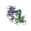
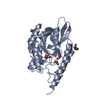
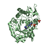
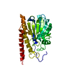
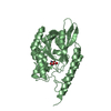
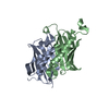
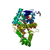
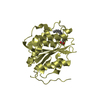
- PDB-6g9z: Crystal structure of human histidine triad nucleotide-binding pro... -

+
Open data


ID or keywords:

Loading...

-
Basic information

Entry
Database: PDB / ID: 6g9z
TitleCrystal structure of human histidine triad nucleotide-binding protein 1 (hHINT1) crystallized at P212121 space group, with visible extended fragment of N-terminus
ComponentsHistidine triad nucleotide-binding protein 1
KeywordsHYDROLASE / phosphoramidase / desulfurase / tumour suppressor
Function / homology
Function and homology information


purine ribonucleotide catabolic process / adenylylsulfatase activity / Hydrolases; Acting on phosphorus-nitrogen bonds / adenosine 5'-monophosphoramidase activity / deSUMOylase activity / protein desumoylation / sulfur compound metabolic process / histone deacetylase complex / intrinsic apoptotic signaling pathway by p53 class mediator / positive regulation of calcium-mediated signaling ...purine ribonucleotide catabolic process / adenylylsulfatase activity / Hydrolases; Acting on phosphorus-nitrogen bonds / adenosine 5'-monophosphoramidase activity / deSUMOylase activity / protein desumoylation / sulfur compound metabolic process / histone deacetylase complex / intrinsic apoptotic signaling pathway by p53 class mediator / positive regulation of calcium-mediated signaling / protein kinase C binding / Hydrolases; Acting on peptide bonds (peptidases); Cysteine endopeptidases / cytoskeleton / hydrolase activity / nucleotide binding / regulation of DNA-templated transcription / signal transduction / proteolysis / extracellular exosome / nucleoplasm / nucleus / plasma membrane / cytoplasm / cytosol
Similarity search - Function
Histidine triad (HIT) protein / HIT domain / Histidine triad, conserved site / HIT domain signature. / HIT domain profile. / HIT-like domain / HIT-like / HIT family, subunit A / HIT-like superfamily / 2-Layer Sandwich / Alpha Beta
Similarity search - Domain/homology
(2S)-2-hydroxybutanedioic acid / Adenosine 5'-monophosphoramidase HINT1
Similarity search - Component
Biological speciesHomo sapiens (human)
MethodX-RAY DIFFRACTION / SYNCHROTRON / MOLECULAR REPLACEMENT / Resolution: 1.43 Å
AuthorsDolot, R.M. / Seda, A. / Nawrot, B.C.
CitationJournal: To Be Published
Title: Differences in crystal packing as the key factor for stabilization of the N-terminal fragment of hHINT1 protein
Authors: Dolot, R.M. / Seda, A. / Nawrot, B.C.
History
DepositionApr 11, 2018Deposition site: PDBE / Processing site: PDBE
SupersessionApr 18, 2018ID: 5O8G
Revision 1.0Apr 18, 2018Provider: repository / Type: Initial release
Revision 1.1Jan 17, 2024Group: Data collection / Database references / Refinement description
Category: chem_comp_atom / chem_comp_bond ...chem_comp_atom / chem_comp_bond / database_2 / pdbx_initial_refinement_model
Item: _database_2.pdbx_DOI / _database_2.pdbx_database_accession

-
Structure visualization

Structure viewerMolecule:
MolmilJmol/JSmol

Downloads & links

-
Assembly

Deposited unit
A: Histidine triad nucleotide-binding protein 1
B: Histidine triad nucleotide-binding protein 1
hetero molecules


Theoretical massNumber of molelcules
Total (without water)27,7823
Polymers27,6482
Non-polymers1341
Water7,224401
1


  • Idetical with deposited unit
  • defined by author&software
  • Evidence: gel filtration
TypeNameSymmetry operationNumber
identity operation1_555x,y,z1
Buried area3840 Å2
ΔGint-17 kcal/mol
Surface area11380 Å2
MethodPISA
Unit cell
Length a, b, c (Å)46.075, 63.379, 76.836
Angle α, β, γ (deg.)90.00, 90.00, 90.00
Int Tables number19
Space group name H-MP212121

-
Components

#1: Protein Histidine triad nucleotide-binding protein 1 / Adenosine 5'-monophosphoramidase / Protein kinase C inhibitor 1 / Protein kinase C-interacting ...Adenosine 5'-monophosphoramidase / Protein kinase C inhibitor 1 / Protein kinase C-interacting protein 1 / PKCI-1


Mass: 13823.931 Da / Num. of mol.: 2
Source method: isolated from a genetically manipulated source
Source: (gene. exp.) Homo sapiens (human) / Gene: HINT1, HINT, PKCI1, PRKCNH1 / Plasmid: pSGA02 / Production host: Escherichia coli BL21 (bacteria) / References: UniProt: P49773, Hydrolases
#2: Chemical ChemComp-LMR / (2S)-2-hydroxybutanedioic acid / L-Malate


Mass: 134.087 Da / Num. of mol.: 1 / Source method: obtained synthetically / Formula: C4H6O5
#3: Water ChemComp-HOH / water


Mass: 18.015 Da / Num. of mol.: 401 / Source method: isolated from a natural source / Formula: H2O

-
Experimental details

-
Experiment

ExperimentMethod: X-RAY DIFFRACTION / Number of used crystals: 1

-
Sample preparation

CrystalDensity Matthews: 2.55 Å3/Da / Density % sol: 51.72 %
Description: Plates of typical dimensions 0.6 x 0.35 x 0.05 mm
Crystal growTemperature: 281 K / Method: vapor diffusion, hanging drop / pH: 7 / Details: 25% PEG 1500, 0.1 M MMT Buffer pH 7.0

-
Data collection

DiffractionMean temperature: 100 K
Diffraction sourceSource: SYNCHROTRON / Site: BESSY / Beamline: 14.1 / Wavelength: 0.918409 Å
DetectorType: DECTRIS PILATUS 6M / Detector: PIXEL / Date: Apr 30, 2016
RadiationMonochromator: Si(111) crystal / Protocol: SINGLE WAVELENGTH / Monochromatic (M) / Laue (L): M / Scattering type: x-ray
Radiation wavelengthWavelength: 0.918409 Å / Relative weight: 1
ReflectionResolution: 1.43→39.52 Å / Num. obs: 40954 / % possible obs: 96.1 % / Redundancy: 3.77 % / Biso Wilson estimate: 15 Å2 / CC1/2: 0.994 / Rmerge(I) obs: 0.137 / Rrim(I) all: 0.161 / Net I/σ(I): 8.78
Reflection shellResolution: 1.43→1.51 Å / Redundancy: 3.74 % / Rmerge(I) obs: 0.703 / Mean I/σ(I) obs: 1.96 / Num. unique obs: 6294 / CC1/2: 0.833 / Rrim(I) all: 0.821 / % possible all: 92.7

-
Processing

Software
NameVersionClassification
REFMAC5.8.0222refinement
XDSdata reduction
XDSdata scaling
MOLREPphasing
RefinementMethod to determine structure: MOLECULAR REPLACEMENT
Starting model: 3TW2
Resolution: 1.43→39.52 Å / Cor.coef. Fo:Fc: 0.857 / Cor.coef. Fo:Fc free: 0.828 / SU B: 4.23 / SU ML: 0.072 / Cross valid method: THROUGHOUT / ESU R: 0.124 / ESU R Free: 0.102 / Details: HYDROGENS HAVE BEEN ADDED IN THE RIDING POSITIONS
RfactorNum. reflection% reflectionSelection details
Rfree0.27726 2041 5 %RANDOM
Rwork0.22362 ---
obs0.2263 38759 95.82 %-
Solvent computationIon probe radii: 0.8 Å / Shrinkage radii: 0.8 Å / VDW probe radii: 1.2 Å
Displacement parametersBiso mean: 9.995 Å2
Baniso -1Baniso -2Baniso -3
1--0.73 Å2-0 Å2-0 Å2
2--0.29 Å2-0 Å2
3---0.44 Å2
Refinement stepCycle: 1 / Resolution: 1.43→39.52 Å
ProteinNucleic acidLigandSolventTotal
Num. atoms1841 0 9 401 2251
Refine LS restraints
Refine-IDTypeDev idealDev ideal targetNumber
X-RAY DIFFRACTIONr_bond_refined_d0.0160.0152069
X-RAY DIFFRACTIONr_bond_other_d0.0010.0171890
X-RAY DIFFRACTIONr_angle_refined_deg1.8121.7472817
X-RAY DIFFRACTIONr_angle_other_deg0.611.7214460
X-RAY DIFFRACTIONr_dihedral_angle_1_deg6.6765276
X-RAY DIFFRACTIONr_dihedral_angle_2_deg36.44219.06264
X-RAY DIFFRACTIONr_dihedral_angle_3_deg14.0915321
X-RAY DIFFRACTIONr_dihedral_angle_4_deg15.4121510
X-RAY DIFFRACTIONr_chiral_restr0.0960.2260
X-RAY DIFFRACTIONr_gen_planes_refined0.010.0212422
X-RAY DIFFRACTIONr_gen_planes_other0.0020.02370
X-RAY DIFFRACTIONr_nbd_refined
X-RAY DIFFRACTIONr_nbd_other
X-RAY DIFFRACTIONr_nbtor_refined
X-RAY DIFFRACTIONr_nbtor_other
X-RAY DIFFRACTIONr_xyhbond_nbd_refined
X-RAY DIFFRACTIONr_xyhbond_nbd_other
X-RAY DIFFRACTIONr_metal_ion_refined
X-RAY DIFFRACTIONr_metal_ion_other
X-RAY DIFFRACTIONr_symmetry_vdw_refined
X-RAY DIFFRACTIONr_symmetry_vdw_other
X-RAY DIFFRACTIONr_symmetry_hbond_refined
X-RAY DIFFRACTIONr_symmetry_hbond_other
X-RAY DIFFRACTIONr_symmetry_metal_ion_refined
X-RAY DIFFRACTIONr_symmetry_metal_ion_other
X-RAY DIFFRACTIONr_mcbond_it2.1180.7251053
X-RAY DIFFRACTIONr_mcbond_other2.1170.7241054
X-RAY DIFFRACTIONr_mcangle_it2.3231.0891346
X-RAY DIFFRACTIONr_mcangle_other2.3231.0891346
X-RAY DIFFRACTIONr_scbond_it1.9270.9051016
X-RAY DIFFRACTIONr_scbond_other1.9280.9031013
X-RAY DIFFRACTIONr_scangle_it
X-RAY DIFFRACTIONr_scangle_other2.5591.2791470
X-RAY DIFFRACTIONr_long_range_B_refined4.38811.5382395
X-RAY DIFFRACTIONr_long_range_B_other3.69810.1142267
X-RAY DIFFRACTIONr_rigid_bond_restr3.45733959
X-RAY DIFFRACTIONr_sphericity_free30.8015213
X-RAY DIFFRACTIONr_sphericity_bonded12.11354085
LS refinement shellResolution: 1.427→1.464 Å / Total num. of bins used: 20
RfactorNum. reflection% reflection
Rfree0.477 146 -
Rwork0.419 2778 -
obs--94.69 %

+
About Yorodumi

-
News

-
Feb 9, 2022. New format data for meta-information of EMDB entries

New format data for meta-information of EMDB entries

  • Version 3 of the EMDB header file is now the official format.
  • The previous official version 1.9 will be removed from the archive.

Related info.:EMDB header

External links:wwPDB to switch to version 3 of the EMDB data model

-
Aug 12, 2020. Covid-19 info

Covid-19 info

URL: https://pdbjlvh1.pdbj.org/emnavi/covid19.php

New page: Covid-19 featured information page in EM Navigator.

Related info.:Covid-19 info / Mar 5, 2020. Novel coronavirus structure data

+
Mar 5, 2020. Novel coronavirus structure data

Novel coronavirus structure data

Related info.:Yorodumi Speices / Aug 12, 2020. Covid-19 info

External links:COVID-19 featured content - PDBj / Molecule of the Month (242):Coronavirus Proteases

+
Jan 31, 2019. EMDB accession codes are about to change! (news from PDBe EMDB page)

EMDB accession codes are about to change! (news from PDBe EMDB page)

  • The allocation of 4 digits for EMDB accession codes will soon come to an end. Whilst these codes will remain in use, new EMDB accession codes will include an additional digit and will expand incrementally as the available range of codes is exhausted. The current 4-digit format prefixed with “EMD-” (i.e. EMD-XXXX) will advance to a 5-digit format (i.e. EMD-XXXXX), and so on. It is currently estimated that the 4-digit codes will be depleted around Spring 2019, at which point the 5-digit format will come into force.
  • The EM Navigator/Yorodumi systems omit the EMD- prefix.

Related info.:Q: What is EMD? / ID/Accession-code notation in Yorodumi/EM Navigator

External links:EMDB Accession Codes are Changing Soon! / Contact to PDBj

+
Jul 12, 2017. Major update of PDB

Major update of PDB

  • wwPDB released updated PDB data conforming to the new PDBx/mmCIF dictionary.
  • This is a major update changing the version number from 4 to 5, and with Remediation, in which all the entries are updated.
  • In this update, many items about electron microscopy experimental information are reorganized (e.g. em_software).
  • Now, EM Navigator and Yorodumi are based on the updated data.

External links:wwPDB Remediation / Enriched Model Files Conforming to OneDep Data Standards Now Available in the PDB FTP Archive

-
Yorodumi

Thousand views of thousand structures

  • Yorodumi is a browser for structure data from EMDB, PDB, SASBDB, etc.
  • This page is also the successor to EM Navigator detail page, and also detail information page/front-end page for Omokage search.
  • The word "yorodu" (or yorozu) is an old Japanese word meaning "ten thousand". "mi" (miru) is to see.

Related info.:EMDB / PDB / SASBDB / Comparison of 3 databanks / Yorodumi Search / Aug 31, 2016. New EM Navigator & Yorodumi / Yorodumi Papers / Jmol/JSmol / Function and homology information / Changes in new EM Navigator and Yorodumi

Read more